
The best meditation for pain relief and stress reduction helps calm the nervous system and improve the way the brain responds to pain, making both physical and emotional discomfort easier to manage. Chronic pain affects more than the body, often leading to anxiety, poor sleep, fatigue, and emotional strain.
Over time, this creates a cycle where stress intensifies pain and pain increases stress. Meditation offers a natural way to break that cycle by promoting relaxation, lowering stress hormones, and improving emotional resilience.
While it is not a replacement for medical treatment, the best meditation for pain relief can become an effective part of a long-term pain management strategy.

How Meditation Helps Reduce Pain and Stress
Meditation reduces pain by calming the brain's response to discomfort while helping the body relax naturally. Pain is more than a physical sensation caused by injured muscles, joints, or nerves.
The brain also plays a major role in determining how strongly pain is felt. When you are stressed, anxious, or emotionally overwhelmed, the brain often amplifies pain signals, making discomfort feel more intense than it actually is.
Regular meditation helps interrupt this cycle by calming the nervous system and encouraging the body to enter a relaxed state. As breathing slows and muscles loosen, the brain becomes less reactive to pain, allowing many people to cope with chronic discomfort more effectively.
Ways Meditation Helps Reduce Pain
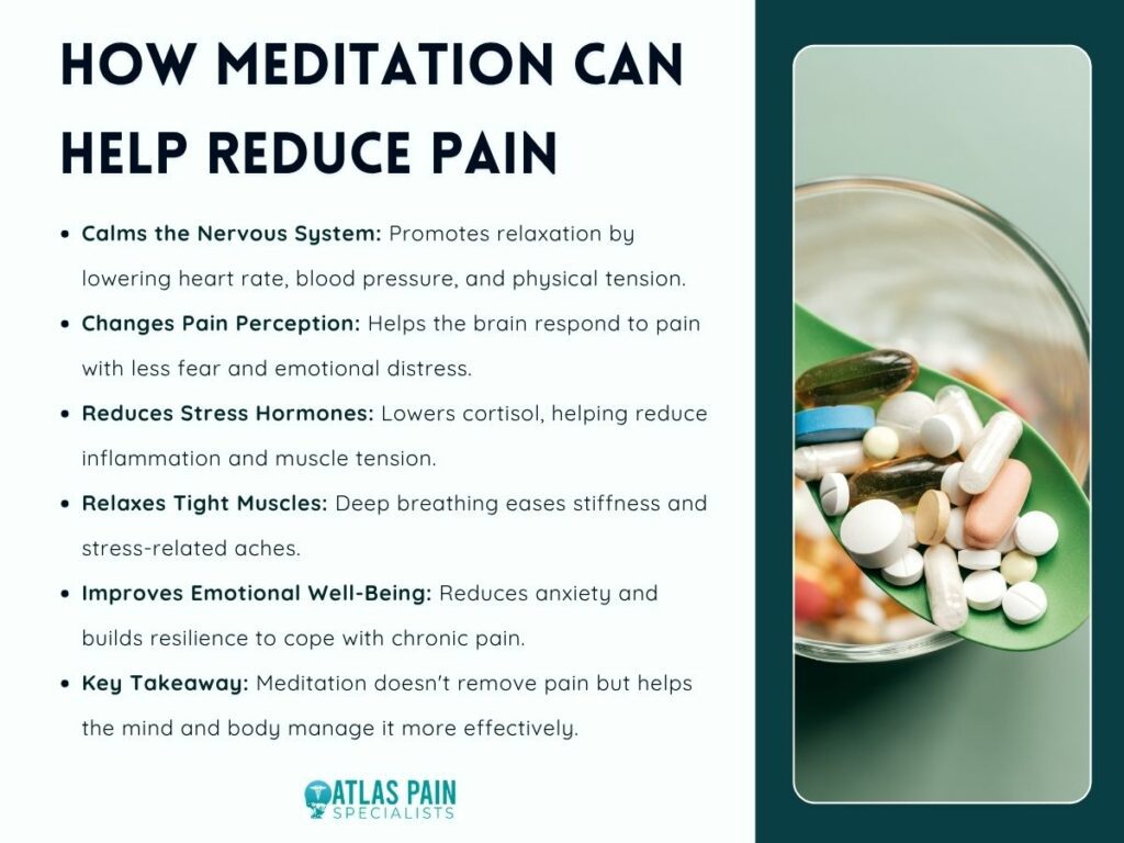
1. Calms the Nervous System
Meditation activates the body's relaxation response, slowing the heart rate, lowering blood pressure, and reducing overall physical tension.
2. Changes How the Brain Processes Pain
With regular practice, meditation helps the brain respond to pain with less fear and emotional distress, making discomfort feel more manageable.
3. Reduces Stress Hormones
Meditation lowers cortisol levels, helping decrease inflammation, muscle tension, and stress that can worsen chronic pain.
4. Relaxes Tight Muscles
Deep breathing and mindful awareness encourage muscles throughout the body to relax, easing stiffness and reducing stress-related aches.
5. Improves Emotional Well-Being
Meditation reduces anxiety and promotes emotional balance, helping people cope better with the mental challenges that often accompany long-term pain.
Meditation does not eliminate the source of pain, but it changes the way the body and mind respond to it. By reducing stress, relaxing muscles, and improving emotional resilience, regular practice can make pain feel less overwhelming and support better overall health.
The Science Behind Meditation and Pain Relief
Research shows that meditation influences the areas of the brain responsible for pain perception and emotional regulation. Scientists once believed meditation worked simply by distracting people from pain.
Today, brain imaging studies tell a different story. Researchers have found that meditation changes activity in brain regions involved in processing pain, attention, and emotions.
These changes help reduce the intensity of pain while improving a person's ability to cope with chronic discomfort. Many healthcare providers now recommend meditation alongside conventional treatments for long-term pain conditions.
Several studies have shown benefits for people living with:
- Chronic lower back pain
- Osteoarthritis
- Rheumatoid arthritis
- Fibromyalgia
- Migraine headaches
- Neuropathic pain
Meditation also improves sleep quality, lowers anxiety, and reduces depression. Since all three strongly influence pain levels, these additional benefits often lead to noticeable improvements in overall quality of life.
Unlike medication, meditation has no significant side effects and can be practiced almost anywhere, making it an accessible tool for long-term pain management.
Best Meditation for Pain Relief Techniques
Different meditation techniques relieve pain in different ways, so choosing the right one depends on your symptoms and personal preference.
Some people prefer quiet mindfulness, while others find guided sessions or breathing exercises easier to follow. There is no single technique that works for everyone, but several approaches have consistently shown positive results.
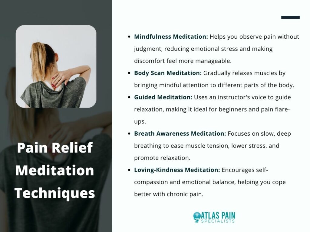
Mindfulness Meditation
Mindfulness meditation is widely considered the best meditation for pain relief because it teaches you to observe pain without immediately reacting to it.
Instead of resisting discomfort, you simply notice it without judgment. This reduces the emotional suffering that often accompanies chronic pain.
A simple mindfulness session usually includes:
- Sitting comfortably
- Focusing on your breathing
- Observing thoughts without following them
- Gently returning attention to your breath
With regular practice, many people notice they become less overwhelmed by pain, even if the physical sensation remains.
Body Scan Meditation
Body scan meditation involves slowly moving your attention through different parts of your body, noticing sensations without trying to change them.
As awareness moves from head to toe, muscles naturally begin to relax. This makes body scans especially useful for people whose pain is linked to muscle tension or widespread discomfort.
Body scan meditation works particularly well for:
- Fibromyalgia
- Chronic neck pain
- Back pain
- Arthritis
- General muscle tension
Many people also find this meditation helpful before bedtime because it encourages deep relaxation.
Guided Meditation
Guided meditation is an excellent option for beginners. Instead of practicing alone, you simply listen to an instructor who leads you through breathing exercises, relaxation techniques, or calming visualizations.
Because someone else provides the guidance, it is often easier to stay focused, especially during painful flare-ups.
Breath Awareness Meditation
Sometimes the simplest meditation techniques are the most effective.
Breath awareness meditation focuses entirely on slow, controlled breathing. Deep breathing activates the body's relaxation response and helps reduce physical tension throughout the body.
Regular breathing meditation may help:
- Lower heart rate
- Relax tight muscles
- Reduce anxiety
- Improve focus
- Decrease stress hormones
Even five minutes of slow breathing can noticeably improve stress-related pain.
Loving-Kindness Meditation
Chronic pain often affects emotional health just as much as physical health.
Loving-kindness meditation focuses on developing compassion toward yourself and others by silently repeating positive phrases such as:
- May I be healthy.
- May I be peaceful.
- May I be free from suffering.
This practice helps reduce frustration, anger, and emotional distress that frequently accompany long-term pain.
Choosing the Best Meditation for Pain Relief Based on Your Condition
Matching your meditation practice to your type of pain often produces better long-term results. Different pain conditions affect the body differently. While every meditation technique promotes relaxation, some are particularly effective for specific health concerns.
| Pain Condition | Best Meditation | Primary Benefit |
| Arthritis | Mindfulness Meditation | Reduces pain sensitivity and stiffness |
| Fibromyalgia | Body Scan Meditation | Encourages full-body relaxation |
| Chronic Back Pain | Guided Meditation | Reduces stress and muscle tension |
| Neck & Shoulder Pain | Breath Awareness | Releases tight muscles |
| Migraines | Mindfulness + Breathing | Lowers stress-related triggers |
| Stress-Related Pain | Loving-Kindness Meditation | Improves emotional well-being |
Although this table provides useful guidance, consistency matters more than choosing the "perfect" technique. The meditation you enjoy practicing regularly is usually the one that produces the greatest benefits.
How to Start a Daily Meditation Practice
A short daily meditation routine is more effective than occasional long sessions. Many beginners think meditation requires an hour of silence every day. In reality, starting with five to ten minutes is often enough to build a lasting habit.
Choose a quiet place where you feel comfortable. Sit in a chair, on a cushion, or even lie down if necessary. Close your eyes and allow your breathing to settle into a slow, natural rhythm.
Instead of trying to empty your mind, simply notice your breathing. Thoughts will come and go, and that is completely normal. Each time your attention wanders, gently bring it back to your breath.
Many people find it easiest to meditate:
- Soon after waking up
- During a lunch break
- After exercise
- Before going to bed
Practicing at the same time each day makes meditation easier to maintain over the long term. As your confidence grows, gradually increase your sessions to fifteen or twenty minutes. Small, consistent practice is far more valuable than occasional lengthy sessions.
Common Challenges and How to Stay Consistent
The biggest obstacle to meditation is expecting perfection instead of steady progress. Starting a meditation practice is simple, but staying consistent can be difficult, especially when you expect immediate results.
Most beginners experience similar challenges, and understanding them can make it easier to build a lasting habit.
Common Challenges Beginners Face
- A Wandering Mind
Many people think they are meditating incorrectly because their thoughts keep drifting. In reality, this is completely normal. Meditation is about gently bringing your attention back to the present each time your mind wanders.
- Physical Discomfort
Sitting for long periods can be uncomfortable, especially if you have chronic pain. Remember that meditation does not require a specific posture. You can sit in a chair, lie down, or use cushions for extra support.
- Expecting Immediate Pain Relief
Some people stop meditating because they do not notice instant improvements. Like exercise, meditation delivers the greatest benefits through regular practice over weeks and months.
- Difficulty Finding Time
Busy schedules often make meditation feel like another task. Starting with just five to ten minutes a day makes it much easier to stay consistent and build the habit.
- Lack of Motivation
It is common to lose motivation, especially during stressful periods. Guided meditation apps, audio sessions, or setting a daily reminder can provide the structure needed to keep practicing.
Although these challenges are common, they become easier to overcome with patience and consistency. Every meditation session, even one that lasts only a few minutes, helps strengthen the habit and supports long-term pain relief, stress reduction, and emotional well-being.
Combining Meditation With Other Pain Management Strategies
The best meditation for pain relief works even better when combined with healthy daily habits and medical care.
Meditation should be viewed as one part of a complete pain management strategy rather than a standalone solution. When combined with other healthy practices, its benefits become even more noticeable.
Many healthcare professionals recommend combining meditation with:
- Regular low-impact exercise
- Physical therapy
- Healthy sleep habits
- Anti-inflammatory nutrition
- Gentle stretching or yoga
- Stress management techniques
- Prescribed medications when needed
These approaches address different aspects of chronic pain. While exercise improves mobility, nutrition reduces inflammation, and physical therapy strengthens muscles, meditation supports the emotional and neurological side of pain management.
Over time, these habits work together to improve physical comfort, emotional resilience, and overall quality of life.
Conclusion
The best meditation for pain relief is the one that fits naturally into your daily life and encourages consistent practice. Whether you choose mindfulness meditation, body scan meditation, guided sessions, breath awareness, or loving-kindness meditation, the goal is the same: helping your body relax while changing the way your mind responds to pain.
Meditation is not an overnight cure, but its benefits grow steadily with time. As stress decreases, sleep improves, and emotional resilience becomes stronger, many people notice that pain feels less overwhelming and easier to manage.
When combined with appropriate medical care and healthy lifestyle habits, meditation becomes a powerful tool for reducing both physical discomfort and everyday stress.
If your shoulder hurts when you lift your arm, it's often due to inflammation, injury, or wear and tear affecting the muscles, tendons, or joints that allow your shoulder to move. Common causes include rotator cuff injuries, shoulder impingement, bursitis, frozen shoulder, tendinitis, and arthritis, all of which can make simple activities like reaching overhead, lifting objects, or getting dressed uncomfortable or painful.
Why does my shoulder hurt when I lift my arm? This article covers the most common causes of shoulder pain when lifting your arm, along with its symptoms, diagnosis, treatment options, prevention tips, and when you should see a healthcare provider.
Understanding How the Shoulder Works
The shoulder is one of the most flexible and mobile joints in the body, allowing you to lift, rotate, push, pull, and reach in almost every direction. This wide range of motion is made possible by a complex network of bones, muscles, tendons, ligaments, cartilage, and fluid-filled sacs that work together to provide strength, stability, and smooth movement. Because so many structures are involved, even a minor injury or inflammation can affect how your shoulder functions and cause pain when lifting your arm.
The main components of the shoulder include:
- The humerus (upper arm bone)
- The scapula (shoulder blade)
- The clavicle (collarbone)
- The rotator cuff muscles, which stabilize and move the shoulder
- Tendons that connect muscles to bones
- Ligaments that hold the bones together
- Bursa sacs that reduce friction between moving structures
- Cartilage that cushions the joint and helps it move smoothly
Since these structures work in a relatively small space, irritation, inflammation, or damage to any one of them can limit movement and make lifting your arm painful or difficult.
Why Does My Shoulder Hurt When I Lift My Arm?
Pain when lifting your arm can occur for many reasons, ranging from minor muscle strains to conditions that affect the shoulder's tendons, joints, or surrounding tissues. The type of pain, where it's located, and when it occurs can help identify the underlying cause.

1. Rotator Cuff Injury
The rotator cuff is a group of four muscles and tendons that stabilize the shoulder and help lift and rotate the arm. When these tendons become inflamed or torn due to overuse, aging, or injury, you may experience pain, weakness, and difficulty raising your arm, especially overhead.
2. Shoulder Impingement Syndrome
Shoulder impingement occurs when the rotator cuff tendons become pinched between the bones of the shoulder during movement. This can lead to pain when lifting your arm, particularly above shoulder height, and may worsen with repetitive overhead activities.
3. Frozen Shoulder (Adhesive Capsulitis)
Frozen shoulder develops when the connective tissue surrounding the shoulder joint thickens and tightens, restricting movement. It usually causes gradually worsening pain and stiffness, making even simple tasks like reaching or dressing difficult.
4. Shoulder Bursitis
Bursitis is the inflammation of the bursa, a small fluid-filled sac that cushions the shoulder joint and reduces friction. An inflamed bursa can make lifting or rotating your arm painful and may also cause tenderness or swelling around the shoulder.
5. Arthritis
Shoulder arthritis occurs when the cartilage that cushions the joint gradually wears away, causing the bones to rub against each other. This can result in persistent pain, stiffness, reduced range of motion, and discomfort during everyday arm movements.
6. Tendinitis
Shoulder tendinitis is caused by inflammation of the tendons, often due to repetitive motions or overuse. The pain typically develops gradually and becomes more noticeable when lifting your arm, reaching overhead, or performing repetitive tasks.
7. Labral Tear
A labral tear affects the ring of cartilage that helps stabilize the shoulder joint. This injury may occur after a fall, heavy lifting, or repetitive overhead movements, leading to pain, clicking sensations, weakness, or a feeling that the shoulder is unstable.
8. Pinched Nerve
Sometimes shoulder pain actually originates from a compressed nerve in the neck rather than the shoulder itself. A pinched nerve may cause pain that radiates into the shoulder and arm, often accompanied by numbness, tingling, or muscle weakness.
While some causes of shoulder pain improve with rest and home care, persistent pain, limited mobility, or significant weakness should be evaluated by a healthcare provider. Identifying the underlying cause early can help ensure appropriate treatment and reduce the risk of long-term shoulder problems.
Common Symptoms That Accompany Shoulder Pain
Shoulder pain when lifting your arm is often accompanied by other symptoms that can provide clues about the underlying condition. The type, location, and severity of these symptoms may vary depending on whether the problem involves the muscles, tendons, joints, bursae, or nerves.
Common symptoms include:
- Sharp pain
- Dull, aching pain
- Burning sensation
- Shoulder stiffness
- Weakness when lifting or carrying objects
- Swelling or tenderness around the joint
- Clicking, popping, or grinding sounds
- Reduced range of motion
- Pain that worsens at night
- Difficulty sleeping on the affected side
Paying attention to these symptoms - including when they occur and what activities make them worse - can help healthcare providers determine the cause of your shoulder pain and recommend the most appropriate treatment.
Risk Factors for Shoulder Pain
While shoulder pain can affect anyone, certain lifestyle habits, occupations, medical conditions, and physical factors can increase your risk of developing shoulder problems. Understanding these risk factors can help you take steps to protect your shoulder health and reduce the likelihood of injury.
These include:
- Aging
- Repetitive overhead work
- Heavy lifting
- Poor posture
- Sports participation, especially those involving overhead movements
- Previous shoulder injury
- Diabetes
- Smoking
- Lack of shoulder strength
- Poor flexibility
People who spend long hours sitting at a desk or using a computer may also develop shoulder pain due to poor posture, prolonged inactivity, and improper workstation ergonomics. Maintaining good posture, taking regular breaks, and strengthening the shoulder muscles can help lower your risk of developing shoulder-related problems.
How Shoulder Pain Is Diagnosed

Diagnosing shoulder pain typically begins with a review of your symptoms, medical history, and a physical examination. Depending on the suspected cause, your healthcare provider may recommend one or more of the following tests:
- Medical history: Your provider will ask about your symptoms, when the pain started, and any previous injuries.
- Physical examination: Your shoulder's range of motion, strength, and areas of tenderness will be evaluated.
- X-rays: Used to detect fractures, arthritis, bone spurs, and other bone-related conditions.
- Ultrasound: Helps identify tendon injuries, bursitis, and rotator cuff problems.
- MRI: Provides detailed images of soft tissues to detect tears, inflammation, and other internal damage.
- CT scan: Offers detailed images of the bones and is useful for evaluating complex injuries.
- Blood tests: May be ordered to check for infections, inflammation, or autoimmune conditions.
An accurate diagnosis helps identify the underlying cause of shoulder pain and guides the most appropriate treatment plan.
Treatment Options for Shoulder Pain
The best treatment for shoulder pain depends on its underlying cause and severity. Many cases improve with conservative treatments, while more serious conditions may require advanced medical care.
- Rest and activity modification: Avoid movements that worsen the pain while allowing the shoulder time to heal.
- Ice therapy: Apply ice packs for 15–20 minutes several times a day to reduce pain and swelling.
- Heat therapy: Use heat to ease muscle tightness and improve flexibility after the initial inflammation has subsided.
- Physical therapy: Strengthens the shoulder muscles, improves flexibility, and restores range of motion.
- Medications: Pain relievers or anti-inflammatory medications can help reduce discomfort and inflammation.
- Corticosteroid injections: May provide temporary relief for severe pain and inflammation when other treatments are not enough.
- Surgery: Recommended for severe injuries, significant tears, or conditions that do not improve with conservative treatment.
Most people recover with nonsurgical treatments, but early diagnosis and following your healthcare provider's recommendations can help speed recovery and prevent further shoulder damage.
Exercises That May Help Improve Shoulder Pain
Once approved by a healthcare professional, gentle exercises can improve flexibility and strength.
Examples include:
- Pendulum swings
- Wall walks
- Cross-body shoulder stretch
- Doorway chest stretch
- External rotation with resistance bands
- Scapular squeezes
Avoid exercises that significantly increase pain, especially during the early stages of recovery.
When Should You See a Doctor?
While mild shoulder pain may improve with rest and home care, some symptoms require prompt medical evaluation. Seek medical attention if you experience any of the following:

1. Pain Lasts Longer Than Two Weeks
Persistent pain that doesn't improve with self-care may indicate an underlying condition that requires treatment. Early evaluation can help prevent the problem from worsening.
2. You Cannot Lift Your Arm
Difficulty or inability to raise your arm could be a sign of a significant tendon injury, frozen shoulder, or another serious condition. A healthcare provider can determine the cause and recommend appropriate treatment.
3. The Shoulder Appears Deformed
A visibly misshapen shoulder may indicate a dislocation or fracture. Seek immediate medical attention, especially if the injury occurred after a fall or accident.
4. Pain Follows a Fall or Injury
Shoulder pain after a traumatic injury may result from fractures, ligament damage, or torn tendons. Prompt evaluation helps ensure proper treatment and recovery.
5. You Experience Numbness or Tingling
Numbness, tingling, or pain that radiates down your arm may indicate nerve involvement. Medical assessment is important to identify and treat the underlying cause.
6. There Is Significant Swelling, Redness, or Fever
These symptoms may be signs of an infection or severe inflammation. Seek medical care as soon as possible for proper diagnosis and treatment.
7. Pain Disrupts Your Sleep or Daily Activities
If shoulder pain regularly interferes with sleep or makes routine tasks difficult, it's time to see a healthcare provider. Persistent symptoms may require targeted treatment to restore function.
Seeking medical care early can help identify the cause of your shoulder pain, relieve symptoms, and reduce the risk of long-term complications.
Can Shoulder Pain Be Prevented?
Although not every shoulder condition is preventable, healthy habits can significantly reduce your risk.
Helpful prevention strategies include:
- Warm up before exercise.
- Strengthen the rotator cuff and upper back muscles.
- Maintain good posture.
- Avoid repetitive overhead movements when possible.
- Use proper lifting techniques.
- Take regular breaks during repetitive work.
- Stretch after workouts.
- Address minor shoulder pain before it becomes chronic.
Building balanced shoulder strength is one of the best long-term protective measures.
Final Thoughts
Experiencing shoulder pain when lifting your arm can make even the simplest daily tasks challenging. Whether the discomfort is caused by an overuse injury, inflammation, or an underlying joint condition, understanding the source of the pain is the first step toward effective treatment and recovery.
Although mild cases may improve with home care, ongoing pain, weakness, or restricted movement should be evaluated by a healthcare provider. With an accurate diagnosis, proper treatment, and preventive measures, you can reduce shoulder pain, regain mobility, and help maintain long-term shoulder health.
Neck pain on the right side is often caused by muscle strain or poor posture, but it can also be a sign of an underlying condition that requires medical attention. Whether the pain develops suddenly after sleeping awkwardly or builds gradually from everyday activities, it can make turning your head, working, and even sleeping uncomfortable.
Because the neck contains muscles, joints, nerves, and spinal structures, identifying the source of the pain is essential for effective treatment. This guide explains the common causes of neck pain on the right side, associated symptoms, treatment options, and the warning signs that mean it's time to see a healthcare provider.
Common Causes of Neck Pain Right Side
Most cases of neck pain on the right side are caused by muscle strain, poor posture, or minor injuries rather than serious disease. Pain on only one side of the neck often develops because the muscles and joints on that side experience more stress than the other.

Sleeping in an awkward position, carrying heavy bags on one shoulder, or spending hours looking down at a phone can overload specific muscles. While many causes are relatively harmless, others involve the spine, nerves, or underlying medical conditions that require treatment.
Muscle Strain
Muscle strain is the leading cause of one-sided neck pain.The muscles that stabilize the neck work continuously throughout the day. Repetitive movements, lifting heavy objects, or sudden twisting can overstretch muscle fibers, leading to inflammation and pain.
Common situations include:
- Sleeping in an unusual position
- Looking down at a laptop for extended periods
- Carrying a backpack on one shoulder
- Sudden exercise without warming up
- Turning the head quickly while driving
The pain usually feels sore, tight, or stiff and often improves within several days with rest.
Poor Posture
Modern lifestyles place tremendous stress on the cervical spine.Hours spent using smartphones, tablets, and computers encourage the head to drift forward.
Every inch the head moves forward increases the amount of pressure placed on the neck muscles. Over time, this continuous strain causes muscle fatigue, stiffness, and localized pain that frequently develops on one side more than the other.
Sleeping Position
The way you sleep significantly affects neck health. Using pillows that are too high or too flat may force your neck into an unnatural position throughout the night.
Similarly, sleeping on your stomach keeps your head turned for several hours, placing uneven stress on the muscles and joints.Many people notice neck pain immediately after waking because these tissues remain irritated after prolonged pressure.
Whiplash and Minor Injuries
Even seemingly minor accidents can injure the neck. Whiplash commonly occurs during rear-end vehicle collisions but may also happen during sports injuries or falls.
The rapid forward and backward movement stretches muscles and ligaments beyond their normal range. Pain may not appear immediately. Instead, stiffness and soreness often develop several hours after the injury.
Medical Conditions That Can Cause Neck Pain Right Side
Although muscle problems are common, persistent neck pain on the right side sometimes results from underlying medical conditions affecting the spine, nerves, or joints.
When pain continues despite rest or gradually worsens, healthcare providers often investigate structural problems within the cervical spine.
Cervical Disc Herniation
The discs between the neck vertebrae act as cushions.If one of these discs bulges or ruptures, it may compress nearby nerves. Depending on which nerve is affected, pain may occur predominantly on the right side of the neck.
Additional symptoms often include:
- Pain radiating into the shoulder
- Arm pain
- Numbness
- Tingling
- Muscle weakness
The pain frequently worsens when coughing, sneezing, or turning the head.
Cervical Arthritis
As people age, the joints in the neck naturally undergo wear and tear. This degeneration can lead to cervical osteoarthritis, causing stiffness, inflammation, and reduced flexibility.
Morning stiffness is particularly common, with symptoms gradually improving as movement increases throughout the day.
Pinched Nerve
A compressed cervical nerve can produce intense one-sided neck pain.Pressure may result from arthritis, disc problems, bone spurs, or swelling around the spinal joints.
Unlike muscle pain, nerve pain often feels sharp, burning, or electric and may extend into the shoulder, arm, or fingers.
Swollen Lymph Nodes
The lymph nodes located along the sides of the neck enlarge when fighting infections.
Common illnesses that may cause swelling include:
- Colds
- Flu
- Ear infections
- Dental infections
- Throat infections
The swollen node may become tender, making the entire right side of the neck feel sore.
Torticollis (Wry Neck)
Torticollis occurs when neck muscles contract involuntarily, forcing the head to tilt toward one side. This condition can develop suddenly after sleeping awkwardly or due to muscle inflammation.
The pain is often severe but usually improves within a few days with appropriate treatment.
Symptoms That Help Identify the Cause
The type of pain and accompanying symptoms often provide valuable clues about what is causing neck pain on the right side. Doctors rarely diagnose neck pain based solely on its location. Instead, they evaluate how the pain behaves, what triggers it, and whether additional symptoms are present.
| Symptom | Possible Cause |
| Dull aching pain | Muscle strain |
| Sharp shooting pain | Pinched nerve |
| Morning stiffness | Arthritis |
| Pain after accident | Whiplash |
| Swollen tender lump | Enlarged lymph node |
| Pain with arm numbness | Herniated disc |
| Muscle tightness | Poor posture |
The duration of symptoms also matters. Pain lasting only a few days often has a different cause than discomfort that continues for several weeks.
When Neck Pain Right Side Becomes a Medical Emergency
Certain symptoms accompanying neck pain on the right side require immediate medical evaluation because they may indicate a serious underlying condition. Although rare, some causes of neck pain involve the spinal cord, brain, blood vessels, or infections that should never be ignored.
Seek emergency care if neck pain occurs together with:
- Sudden weakness in an arm or leg
- Loss of bladder or bowel control
- Difficulty walking
- High fever with severe neck stiffness
- Confusion
- Difficulty speaking
- Severe headache unlike previous headaches
- Chest pain
- Trouble breathing
- Loss of consciousness
Similarly, neck pain following a major car accident or fall should always be evaluated, even if symptoms initially appear mild.
How Neck Pain Right Side Is Diagnosed
Accurate diagnosis begins with a detailed medical history and physical examination rather than immediate imaging tests. Most healthcare providers first ask when the pain began, whether an injury occurred, what movements worsen symptoms, and whether numbness or weakness is present.
During the examination, they evaluate:
- Neck movement
- Muscle tenderness
- Reflexes
- Arm strength
- Sensation
- Range of motion
If symptoms suggest a more complex condition, additional tests may be recommended.
Diagnostic Tests
Depending on the suspected cause, your healthcare provider may order:
| Test | Purpose |
| X-ray | Evaluates bones and arthritis |
| MRI | Detects disc problems and nerve compression |
| CT scan | Provides detailed bone images |
| Electromyography (EMG) | Assesses nerve function |
| Blood tests | Detect infection or inflammatory disease |
Imaging is generally reserved for persistent symptoms, neurological deficits, or suspected serious conditions.
Treatment Options for Neck Pain Right Side
Treatment depends on the underlying cause, but most people recover successfully with conservative care. Many cases improve within one to three weeks without surgery. The primary goals are reducing pain, restoring mobility, and preventing recurrence.

Home Care
For recent muscle-related pain, simple treatments are often highly effective.
Recommended strategies include:
- Rest from aggravating activities
- Gentle stretching
- Ice during the first 48 hours
- Heat after initial inflammation subsides
- Maintaining good posture
- Using supportive pillows
Remaining completely inactive is generally discouraged because gentle movement helps prevent stiffness.
Medications
Over-the-counter medications may help reduce discomfort. Depending on the cause, healthcare providers may recommend:
- Acetaminophen
- Nonsteroidal anti-inflammatory drugs (NSAIDs)
- Muscle relaxants
- Prescription anti-inflammatory medications
Medication works best when combined with posture correction and activity modification rather than used alone.
Physical Therapy
Physical therapy is especially valuable for recurring neck pain. A therapist develops individualized exercises that strengthen the muscles supporting the cervical spine while improving flexibility and posture.
Treatment may include manual therapy, stretching, strengthening exercises, ergonomic education, and mobility training.
Injections and Advanced Treatments
Persistent nerve-related pain sometimes requires additional treatment. Options may include corticosteroid injections around irritated nerves to reduce inflammation and provide temporary relief while rehabilitation continues.
Surgery is considered only when conservative treatment fails or when significant nerve compression causes progressive weakness or spinal cord involvement.
Preventing Neck Pain Right Side From Returning
Most episodes of neck pain on the right side can be prevented by reducing everyday strain on the neck muscles and joints. Once symptoms improve, prevention becomes just as important as treatment.
Many people unknowingly continue habits that place repeated stress on the cervical spine, leading to recurring discomfort.
A few consistent lifestyle changes can make a significant difference:
- Maintain good posture while sitting and standing.
- Position computer screens at eye level.
- Take regular breaks during desk work.
- Avoid cradling the phone between your shoulder and ear.
- Use a pillow that keeps your neck in a neutral position.
- Strengthen your neck, shoulder, and upper back muscles.
- Stretch throughout the day if you spend long hours sitting.
Even small adjustments, such as raising a laptop screen or switching shoulders when carrying a bag, reduce uneven stress on the neck over time.
People who exercise regularly should also pay attention to technique. Lifting weights with poor form or performing repetitive overhead movements can place unnecessary strain on the neck and shoulders.
Warming up before activity and maintaining proper form lowers the risk of injury.
When to See a Doctor for Neck Pain Right Side
While many cases improve within a few days, persistent or worsening neck pain right side should be evaluated by a healthcare professional.
Schedule a medical appointment if your pain:
- Lasts longer than one to two weeks
- Continues to worsen despite home treatment
- Frequently returns
- Limits your ability to work or sleep
- Spreads into the shoulder or arm
- Causes numbness, tingling, or weakness
- Develops after a fall or accident
Early evaluation helps identify problems before they become more difficult to treat. Conditions such as cervical disc herniation, arthritis, or nerve compression often respond better when managed promptly rather than after symptoms have progressed.
Conclusion
Neck pain on the right side is often linked to everyday issues such as muscle strain, poor posture, or sleeping in an awkward position. In many cases, these problems improve with rest, gentle movement, and simple lifestyle adjustments.
However, persistent pain, recurring episodes, or symptoms that radiate into the arm may point to conditions involving the cervical spine, nerves, or joints that deserve medical attention. Understanding what neck pain on the right side means allows you to respond appropriately instead of simply masking the discomfort.
Paying attention to accompanying symptoms, addressing posture and ergonomic habits, and seeking timely medical care when needed can help prevent long-term complications and support a faster recovery.
A clicking sound from the knee during walking often prompts concern about joint integrity. The noise can originate from mechanical or physiological processes within the femur, tibia, and patella.
Why Does My Knee Click When I Walk? In many cases, this phenomenon results from harmless events like gas release from synovial fluid or tendon snapping over bone. These instances do not indicate structural damage or functional limitation.
However, a click that repeats with each step and occurs with discomfort or swelling warrants clinical assessment. Such presentations may signal meniscal tears, chondromalacia, or early degenerative changes requiring evaluation.
The Basic Anatomy of the Knee
The knee functions as a modified hinge joint that permits flexion and extension along with a small degree of rotation. Its structural integrity depends on the interaction between bony surfaces, articular cartilage, menisci, ligaments, and the surrounding musculature.
Bony Structures
The primary bones that form the knee joint include the distal femur, the proximal tibia, and the patella. The femur and tibia articulate through their condyles, while the patella sits within the trochlear groove of the femur.
The patella serves as a sesamoid bone that increases the mechanical advantage of the quadriceps tendon during extension. This bony arrangement creates the foundational framework for all subsequent movement and load transmission.
Cartilage and Menisci
Articular cartilage covers the ends of the femur, tibia, and the posterior surface of the patella. This hyaline cartilage provides a smooth, low-friction surface that permits joint movement with minimal resistance.
Two C-shaped fibrocartilaginous structures, the medial and lateral menisci, sit between the femoral condyles and the tibial plateau. These menisci act as shock absorbers and improve joint congruity during weight-bearing activities.
Synovial Fluid and Joint Capsule
A fibrous joint capsule encloses the knee and contains the synovial membrane. The synovial membrane produces synovial fluid, which lubricates the articular surfaces and nourishes the avascular cartilage.
Synovial fluid contains dissolved gases that can form bubbles under changes in joint pressure. This fluid also reduces friction between the moving components during gait.
Ligaments and Tendons
The major ligaments of the knee include the anterior cruciate, posterior cruciate, medial collateral, and lateral collateral ligaments. These structures provide passive stability by restricting excessive anterior, posterior, and rotational movements.
The quadriceps tendon connects the quadriceps muscle to the patella, and the patellar tendon continues from the patella to the tibial tuberosity. These tendons transmit force from the thigh muscles to produce knee extension and control flexion during walking.
The Benign and Harmless Reasons for Clicking (Crepitus)
Crepitus is the medical term for the audible or palpable clicking, popping, or grating sensation that occurs during joint movement. This phenomenon is frequently observed in the knee and does not necessarily indicate pathology.

Several harmless physiological processes can produce these sounds without causing tissue damage or pain. Distinguishing these benign causes from pathological ones requires attention to accompanying symptoms.
Gas Bubble Formation
Synovial fluid contains dissolved nitrogen, oxygen, and carbon dioxide. A sudden change in joint pressure during movement can cause these gases to form microscopic bubbles that subsequently collapse or burst.
This process, known as cavitation, produces a sharp popping sound similar to that heard during knuckle cracking. The bubbles reform within the fluid over time and the sound can recur with normal joint motion.
Tendon or Ligament Snapping
The iliotibial band and the biceps femoris tendon cross over the bony prominences of the femur and tibia. During flexion and extension of the knee, these structures can shift position and snap over the condyles or fibular head.
This snapping occurs more frequently when the tendons are tight or when the joint moves through a specific range of motion. The sound is typically crisp and singular, without associated pain or inflammation.
Surface Irregularities
The articular cartilage surface can develop minor roughness or fibrillation as a normal part of aging. When the femoral condyles glide over the tibial plateau, these microscopic irregularities can create a grating or clicking noise.
This type of crepitus is often positional and may vary with changes in activity level or joint loading. The absence of pain, swelling, or mechanical symptoms suggests a benign process related to normal wear.
When the Clicking is Accompanied by Pain (Potential Causes)
A click that occurs with each step and is accompanied by pain requires a different clinical consideration than a silent pop. The presence of discomfort, swelling, or mechanical interference suggests an underlying structural or degenerative condition within the joint.
Several specific pathologies can produce an audible click alongside pain during ambulation. Each condition presents with distinct characteristics that help guide clinical assessment.
Torn Meniscus
A meniscal tear often produces a distinct click or pop at the moment of injury or during subsequent weight-bearing movements. The torn fragment can displace into the joint space and create a mechanical block that produces an audible sound with each step.
This clicking is frequently associated with joint line tenderness, localized swelling, and a sensation of catching or locking. The pain typically worsens with twisting motions or squatting and may limit full knee extension.
Chondromalacia Patella
Chondromalacia refers to the softening and deterioration of the cartilage on the undersurface of the patella. This condition causes a grinding or clicking sensation that is most noticeable during stair climbing or when rising from a seated position.
The pain is typically located behind or around the kneecap and intensifies with prolonged sitting or increased activity. The crepitus arises from the irregular patellar surface tracking against the femoral trochlea during knee flexion.
Osteoarthritis
Osteoarthritis involves progressive loss of articular cartilage and subsequent narrowing of the joint space. The exposed subchondral bone surfaces can rub against each other during movement and produce coarse crepitus.
This clicking is often accompanied by morning stiffness that improves with activity and deep aching pain that worsens with weight-bearing. The condition develops gradually over years and is more prevalent in older adults and those with a history of joint trauma.
Loose Body
A loose body consists of a fragment of bone, cartilage, or osteophyte that has detached and floats freely within the joint cavity. This fragment can move into the joint space during walking and produce a sudden click or catch.
The patient may experience intermittent locking where the knee cannot fully extend or flex. The symptoms are often unpredictable and vary depending on the location of the loose fragment at any given moment.
Other Symptoms to Watch for Alongside the Click
The click itself is less clinically significant than the symptoms that accompany it. A thorough symptom inventory helps differentiate benign crepitus from conditions that require intervention.
Several concurrent findings should prompt a more detailed clinical evaluation. These symptoms indicate that the clicking is not an isolated event but part of a broader joint process.
Swelling and Inflammation
Joint effusion, or visible swelling, often develops in response to irritation or injury within the knee. This fluid accumulation can stretch the joint capsule and contribute to stiffness and discomfort.
The presence of warmth or redness over the joint suggests an inflammatory component. Swelling that occurs within hours of activity may indicate acute injury, whereas gradual swelling points toward degenerative or chronic conditions.
Instability or Giving Way
A sensation that the knee will buckle or collapse during weight-bearing is a concerning symptom. This instability often results from ligamentous laxity or muscle weakness that compromises joint control.
The feeling of giving way can occur with or without pain and may increase the risk of falls. This symptom warrants prompt assessment to determine the underlying cause and prevent further joint damage.
Locking or Catching
The knee may fail to fully extend or flex during movement, a phenomenon described as locking. True mechanical locking typically indicates a displaced meniscal fragment or loose body that physically blocks joint motion.
Pseudo-locking, in contrast, occurs when pain or muscle spasm prevents full range of motion. Both forms of locking interfere with normal gait and require careful evaluation to distinguish their origins.
Persistent or Sharp Pain
Pain that remains constant or intensifies with each step is a primary indicator of joint pathology. Sharp, stabbing sensations often correlate with acute meniscal tears or impingement of soft tissues.
Dull, aching pain that worsens after activity and improves with rest is more consistent with degenerative arthritis. The character, location, and timing of the pain provide essential diagnostic clues.
Common Treatment Options
The treatment approach for a clicking knee depends entirely on the underlying diagnosis established through clinical examination and imaging. Management strategies range from conservative rehabilitation to surgical intervention based on the specific pathology.
Many conditions respond well to non-operative measures that address muscle strength, joint mechanics, and inflammation. Surgical options are reserved for cases where structural abnormalities cause persistent mechanical symptoms or functional impairment.
Physical Therapy
Therapeutic exercise programs focus on strengthening the quadriceps, hamstrings, and hip musculature to improve joint stability. These muscles provide dynamic support that reduces abnormal tracking and excessive loading of articular surfaces.
Stretching exercises address tightness in the iliotibial band and hamstrings, which can alter patellar mechanics. A tailored rehabilitation protocol can reduce symptoms in many patients within several weeks.
Pharmacologic Management
Non-steroidal anti-inflammatory drugs reduce pain and inflammation associated with osteoarthritis and soft tissue irritation. These medications are used on a short-term basis to facilitate participation in physical therapy.
Topical preparations such as diclofenac gel provide local relief with minimal systemic side effects. Oral analgesics like acetaminophen may be sufficient for patients with mild to moderate discomfort.
Corticosteroid Injections
Intra-articular corticosteroid injections deliver high-concentration anti-inflammatory medication directly into the joint space. These injections can provide significant pain relief for patients with osteoarthritis or inflammatory conditions.
The therapeutic effect typically lasts for several weeks to months. Repeat injections are performed with caution to avoid potential cartilage damage from cumulative steroid exposure.
Viscosupplementation
Hyaluronic acid injections supplement the natural synovial fluid in patients with knee osteoarthritis. This treatment aims to improve joint lubrication and reduce friction during movement.
The clinical benefit varies among patients and may not be superior to corticosteroid injections in all cases. This option is generally considered after conservative measures have failed to provide adequate relief.
Surgical Intervention
Arthroscopic surgery allows visualization and treatment of intra-articular pathology through small incisions. Common procedures include partial meniscectomy for unstable meniscal tears and removal of loose bodies.
Osteotomy realigns the mechanical axis of the lower extremity to offload damaged joint compartments. Total knee arthroplasty is reserved for end-stage osteoarthritis with severe pain and functional limitation.
Conclusion
A clicking sound during walking is often a benign phenomenon that requires no intervention. The absence of pain, swelling, or functional limitation suggests a harmless mechanical process.
Painful clicking with associated symptoms indicates the need for a systematic evaluation. Clinical examination and imaging studies can identify the specific pathology and guide appropriate management.
Conservative measures such as activity modification and physical therapy address many common conditions. Surgical intervention remains an option for structural problems that do not respond to non-operative treatment.
Shoulder pain from sleeping on your side can be caused by prolonged pressure on the shoulder joint, rotator cuff irritation, bursitis, shoulder impingement, poor alignment, or underlying conditions that affect the surrounding muscles and tissues. Spending hours in the same sleeping position may place excess stress on the shoulder, leading to pain, stiffness, and discomfort upon waking.
This article examines the possible causes of shoulder pain from sleeping on side. It includes associated risk factors, and symptoms that may require medical attention. It also covers treatment options, daytime physical therapy and preventive exercises, sleep position adjustments, and tips for choosing the best pillow for side sleepers.
Why Sleeping on Your Side Can Cause Shoulder Pain
Sleeping on your side is a common and comfortable position for many people, but it can place sustained pressure on the shoulder joint and surrounding tissues. Over time, this pressure may lead to irritation, inflammation, and discomfort, especially if proper support and alignment are lacking.
1. Increased Pressure on the Shoulder Joint
Sleeping on one side for several hours places much of your upper body weight directly on the shoulder joint. This constant pressure can compress soft tissues, reduce circulation, and contribute to soreness or stiffness upon waking.
2. Rotator Cuff Irritation
The rotator cuff muscles and tendons help stabilize the shoulder and support arm movement. Prolonged compression during sleep may irritate these structures, causing pain, tenderness, and discomfort that often becomes more noticeable at night.
3. Shoulder Impingement
Shoulder impingement occurs when tendons are pinched or compressed within the shoulder joint. Sleeping on the affected side can worsen this compression, leading to pain, reduced mobility, and difficulty finding a comfortable sleeping position.
4. Bursitis
Bursae are small fluid-filled sacs that cushion the shoulder and reduce friction between tissues. Continuous pressure from side sleeping can irritate these sacs, resulting in inflammation, tenderness, and aching that may persist throughout the day.
5. Poor Sleeping Alignment
Using an unsupportive pillow or sleeping in an awkward position can place excess strain on the shoulder, neck, and upper back. Poor alignment may increase muscle tension and joint stress, contributing to recurring shoulder pain over time.
6. Underlying Shoulder Conditions
Pre-existing conditions such as arthritis, tendinitis, frozen shoulder, or previous injuries can make the shoulder more sensitive to pressure during sleep. Side sleeping may aggravate these issues and intensify symptoms, particularly in individuals with chronic shoulder problems.
Although occasional shoulder discomfort from side sleeping is common, persistent or worsening pain may indicate an underlying condition that requires attention. Improving sleep posture, using proper support, and addressing existing shoulder issues can help reduce pain and promote more restful sleep.
Other Factors That May Contribute to Shoulder Pain
In addition to sleeping position, several underlying conditions and lifestyle factors can increase the risk of shoulder pain. These issues may make the shoulder more vulnerable to discomfort during sleep.
1. Previous Shoulder Injuries
Old injuries can weaken shoulder structures and make them more sensitive to pressure. Pain may return or worsen when sleeping on the affected side.
2. Arthritis
Arthritis causes joint inflammation and stiffness that can intensify during periods of inactivity. Sleeping on the shoulder may increase discomfort.
3. Tendinitis
Inflamed tendons are more susceptible to irritation from prolonged pressure. This can lead to pain that is especially noticeable at night.
4. Frozen Shoulder
Frozen shoulder restricts movement and causes persistent pain. Side sleeping may aggravate symptoms and reduce sleep quality.
5. Muscle Imbalances
Weak or tight muscles can alter shoulder mechanics and create added strain. This may contribute to pain and stiffness over time.
6. Age-Related Wear and Tear
Natural changes in joints and soft tissues can increase shoulder sensitivity. Older adults may be more likely to experience discomfort from side sleeping.
Identifying these contributing factors can help address the root cause of shoulder pain. Managing underlying conditions may improve comfort and support better sleep.
How to Prevent Shoulder Pain While Sleeping on Your Side
Making a few adjustments to your sleep habits can help reduce pressure on the shoulder and improve overall comfort. Proper support and positioning are key to preventing nighttime pain.
1. Use the Right Pillow
Choose a pillow that keeps your head, neck, and spine properly aligned. Adequate support helps minimize strain on the shoulder joint.
2. Hug a Pillow
Holding a pillow against your chest can support the upper arm and relieve pressure on the shoulder. This position may also improve body alignment.
3. Choose an Appropriate Mattress
A medium-firm mattress can provide a balance of comfort and support. It helps distribute body weight more evenly and reduces pressure points.
4. Alternate Sleeping Positions
Changing positions during the night can prevent prolonged pressure on one shoulder. Sleeping on your back occasionally may also provide relief.
5. Stretch Regularly
Gentle stretching exercises can improve flexibility and reduce muscle tension. Regular movement may help keep the shoulders healthy and mobile.
Simple changes to your sleep setup and daily routine can make a noticeable difference in shoulder comfort. Consistently practicing these habits may help prevent pain and promote better sleep quality.
When Shoulder Pain Could Signal a More Serious Problem
Occasional discomfort after sleeping awkwardly is common. However, certain symptoms may indicate a condition that requires medical attention.
Consult a healthcare provider if you experience:
- Severe or worsening pain
- Persistent pain lasting several weeks
- Significant weakness
- Limited shoulder movement
- Swelling or redness
- Pain following an injury
- Numbness extending into the arm or hand
Early diagnosis and treatment can help prevent minor issues from progressing into chronic conditions and support a faster recovery.
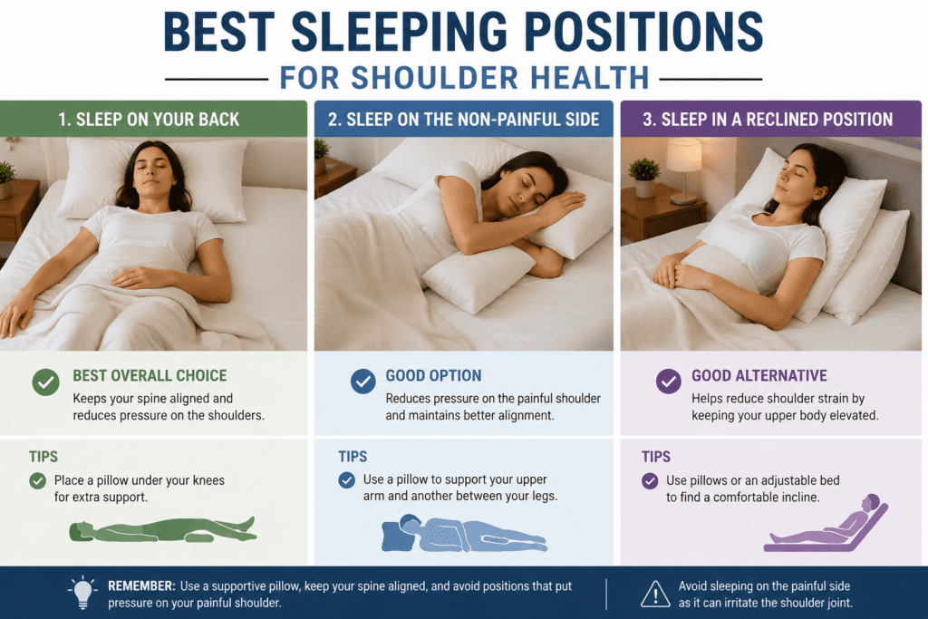
Best Sleeping Positions for Shoulder Health
The way you sleep can significantly affect shoulder comfort and overall joint health. Choosing supportive sleeping positions may help reduce pressure, improve alignment, and minimize nighttime pain.
1. Sleeping on Your Back
Sleeping on your back distributes body weight evenly and places less pressure on the shoulders. This position also helps maintain proper spinal alignment.
2. Side Sleeping with Support
Placing a pillow between your arms or hugging a pillow can help support the upper shoulder and reduce strain. It also promotes better alignment of the neck, shoulders, and spine.
3. Elevated Sleeping Position
Slightly elevating your upper body with pillows may help reduce pressure and inflammation in the shoulder joint. This position can be especially beneficial for people with shoulder impingement or bursitis.
Adopting a shoulder-friendly sleeping position can improve comfort and support better sleep quality. Combining proper positioning with supportive pillows and mattresses may further reduce the risk of shoulder pain.
How to Choose the Perfect Pillow for Side Sleepers
Your pillow can significantly impact shoulder health, especially for side sleepers. A pillow that is too high or too low may force the neck out of alignment, placing additional strain on the shoulders, surrounding muscles, and nerves extending into the arm.
| Pillow Characteristic | Recommended for Side Sleepers | Why It Matters |
| Pillow Loft (Height) | High loft (approximately 4–6 inches) | The pillow should fill the space between the neck and shoulder to help maintain proper spinal alignment throughout the night. |
| Material Firmness | Medium-firm materials such as memory foam or latex | Pillows that are too soft can flatten under the head's weight, causing the neck to tilt and increasing pressure on the shoulder joint. |
| Contour Design | Ergonomic or contoured pillows | Contoured designs support the natural curve of the neck and may help reduce stress on the shoulders and surrounding nerves. |
The ideal pillow should keep your head, neck, and spine in a neutral position while minimizing pressure on the shoulder. Choosing the right combination of height, firmness, and support can improve sleep quality and help prevent shoulder discomfort over time.
Daytime Physical Therapy and Preventive Exercises
Incorporating simple exercises into your daily routine can help strengthen the shoulder, improve mobility, and reduce discomfort caused by side sleeping. Regular movement may also prevent stiffness and support long-term shoulder health.
1. Pendulum Swings
This gentle exercise helps increase shoulder mobility and reduce joint stiffness. It is often recommended for easing mild shoulder discomfort.
2. Cross-Body Stretch
Bringing one arm across the chest helps stretch the muscles around the shoulder. This movement can improve flexibility and relieve tension.
3. Doorway Chest Stretch
Stretching the chest muscles may help improve posture and reduce strain on the shoulders. It can also counteract the effects of prolonged sitting.
4. Shoulder Blade Squeezes
Pinching the shoulder blades together strengthens the upper back muscles and promotes better alignment. This exercise may help reduce stress on the shoulder joint.
5. Resistance Band Rotations
Light resistance exercises can strengthen the rotator cuff muscles and improve shoulder stability. They may also help lower the risk of future injuries.
6. Posture Awareness Exercises
Practicing proper posture throughout the day helps keep the shoulders in a healthier position. Good alignment can reduce unnecessary pressure and muscle tension.
Performing these exercises consistently can improve shoulder function and support better sleep comfort. If pain persists or worsens, consult a healthcare professional or physical therapist for personalized guidance.
Simple Lifestyle Habits to Support Shoulder Health
Daily habits can affect shoulder comfort and may help prevent recurring pain. Small adjustments throughout the day can reduce strain and support joint health.
- Take Regular Movement Breaks - Avoid staying in one position for extended periods. Gentle movement helps reduce stiffness and maintain flexibility.
- Maintain Good Posture - Proper posture keeps the shoulders aligned and reduces unnecessary stress on the joint.
- Avoid Carrying Heavy Bags on One Side - Repeatedly carrying heavy loads on one shoulder can increase strain and contribute to discomfort.
- Stay Physically Active - Regular exercise strengthens the muscles that support the shoulders and promotes better mobility.
Simple lifestyle changes can complement healthy sleep habits and preventive exercises. Over time, they may help reduce shoulder pain and improve overall shoulder health.
Final Thoughts
Shoulder pain caused by sleeping on your side is commonly linked to prolonged pressure on the joint, poor sleeping posture, or pre-existing shoulder issues. Understanding the underlying cause and making targeted changes to your sleep setup can help ease discomfort and promote more restful sleep.
Choosing the right pillow, improving sleep positioning, and staying active with regular stretching and strengthening exercises can support long-term shoulder health. However, persistent or worsening pain should be evaluated by a healthcare professional to identify any underlying conditions and determine the most appropriate treatment approach.
Lower back pain during standing presents a common clinical complaint that often stems from mechanical stress on the spinal column. The pain emerges from a complex interaction between muscle endurance, joint integrity, and neural structures.
Many individuals find that what begins as a mild ache gradually intensifies the longer they remain upright. This progression provides a critical diagnostic clue for healthcare providers.
The causes range from simple muscle fatigue to degenerative conditions of the spine. Each of the eight distinct mechanisms discussed here offers a pathway toward targeted treatment and prevention strategies. Let's look at 8 causes of lower back pain when standing.
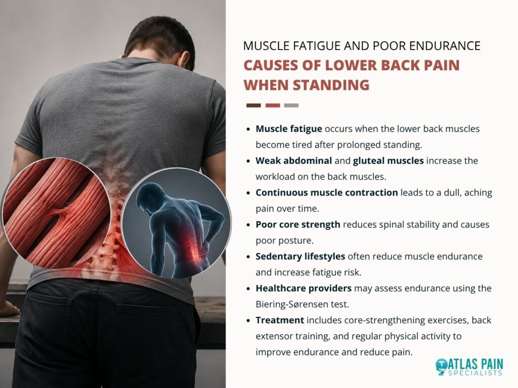
1. Muscle Fatigue and Poor Endurance
Sustained standing forces the paraspinal muscles to contract continuously against gravity. Weakness in the abdominal and gluteal muscles compounds this workload because the erector spinae must compensate for insufficient core support.
Fatigue Onset and Core Instability
The first sign of muscle fatigue is often a dull ache that worsens over time. This discomfort develops as metabolic byproducts accumulate within the muscle tissue.
Core musculature functions as a natural corset that stabilizes the lumbar spine. Inadequate core strength permits excessive anterior pelvic tilt during standing, which shifts the center of gravity forward and forces the lower back muscles to work harder.
Endurance and Clinical Assessment
Low endurance in the back extensors predicts earlier onset of standing-related pain. Individuals with sedentary occupations often display reduced paraspinal endurance and remain vulnerable to discomfort during routine standing tasks.
Healthcare providers evaluate muscle fatigue through endurance tests such as the Biering-Sørensen test. A short hold time indicates poor extensor endurance and suggests a need for targeted strengthening.
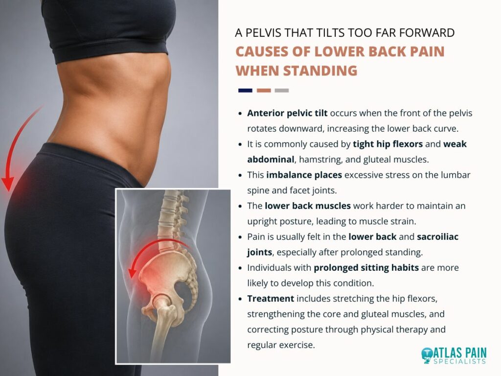
2. A Pelvis That Tilts Too Far Forward
Anterior pelvic tilt describes a postural deviation where the front of the pelvis drops downward and the rear elevates. This position increases the lumbar lordosis curve and places excessive compression on the facet joints of the lower spine.
The muscle imbalance responsible for this tilt involves tight hip flexors and weak hamstrings. The erector spinae muscles contract forcefully to counterbalance the forward pull of the pelvis, which leads to chronic strain.
The Postural Mechanism
The iliopsoas muscle shortens in individuals who sit for prolonged periods. This shortened position pulls the pelvis into an anterior tilt whenever the person stands.
The abdominal muscles become lengthened and weakened in this posture. Their reduced ability to oppose the hip flexors permits the tilt to persist throughout standing activities.
Pain Distribution and Symptoms
Pain from anterior pelvic tilt localizes to the lower lumbar region and the sacroiliac joints. The ache often presents as a deep, diffuse discomfort rather than a sharp, localized sensation.
Patients frequently report increased pain at the end of the day. This pattern reflects the cumulative loading on spinal structures over hours of standing.
Correction Strategies
Stretching the hip flexors and quadriceps reduces the anterior pull on the pelvis. Strengthening the gluteal muscles provides the posterior counterforce needed to restore neutral alignment.
A physical therapist can assess pelvic position during standing and sitting. Treatment typically includes a home exercise program focused on these specific muscle groups.
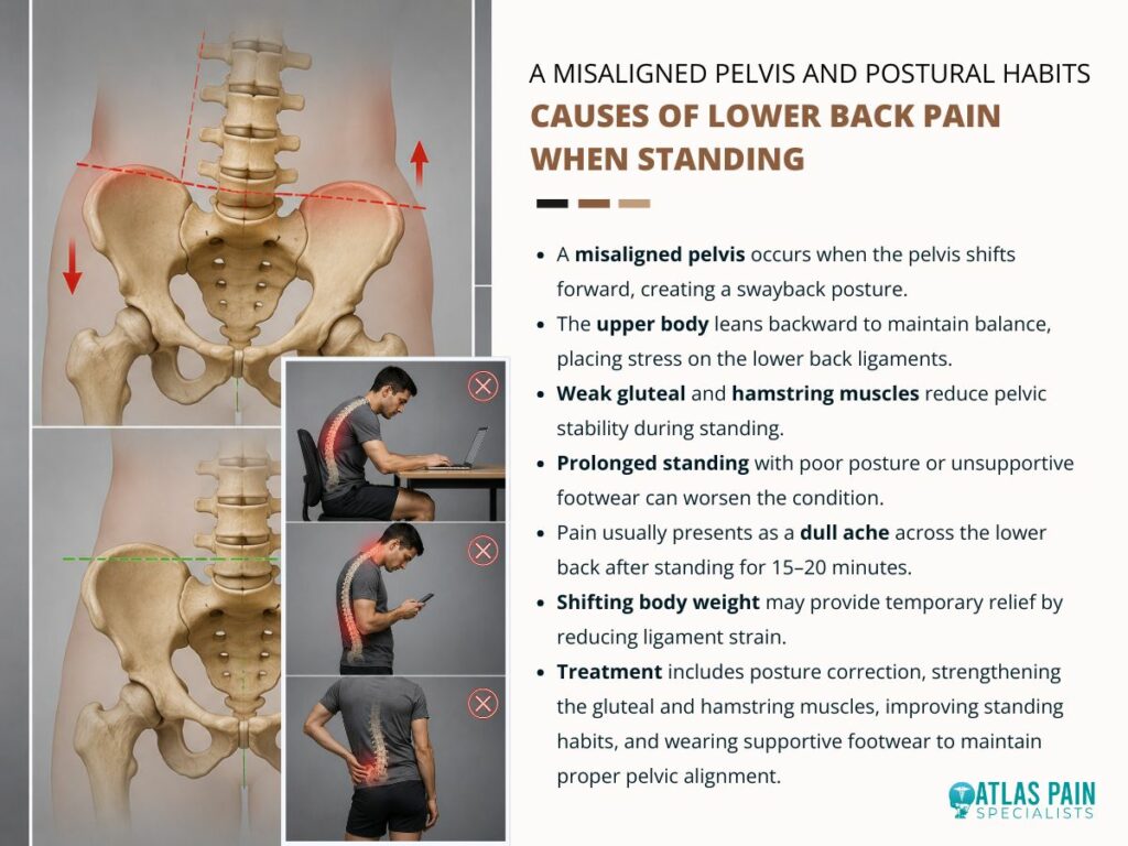
3. A Misaligned Pelvis and Postural Habits
Swayback posture differs from anterior pelvic tilt in that the entire pelvis shifts forward relative to the feet. The upper body leans backward to maintain balance, which creates a straight but slouched spinal alignment.
This posture places excessive stress on the posterior ligaments of the lumbar spine. The ligaments stretch over time and lose their ability to provide passive support during standing.
The Swayback Mechanism
The hip joints extend and the thoracic spine moves posteriorly in this stance. The lower back flattens rather than curves excessively, which redistributes load to the connective tissues.
The hamstrings and gluteal muscles remain in a lengthened and weakened state. Their reduced activation forces the lumbar ligaments and joint capsules to bear more of the gravitational load.
Pain Characteristics
Pain from swayback posture typically presents as a chronic ache across the lower back. The discomfort intensifies after fifteen to twenty minutes of quiet standing.
Patients often report that shifting weight from one foot to the other provides temporary relief. This movement reduces ligamentous tension and redistributes compressive forces across the spine.
Habitual Factors and Correction
Prolonged standing on soft surfaces or with poor footwear contributes to this postural pattern. The lack of proprioceptive feedback from the feet allows the pelvis to drift forward without conscious correction.
Re-education of standing posture focuses on weight distribution over the midfoot. Training the gluteal muscles to engage during standing helps anchor the pelvis in a more stable position.
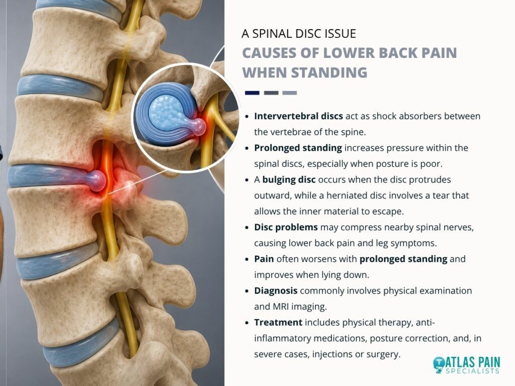
4. A Spinal Disc Issue
Intervertebral discs serve as shock absorbers between the vertebral bodies. Each disc contains a gelatinous nucleus pulposus surrounded by a tough annulus fibrosus.
Standing increases the hydrostatic pressure within the disc's nucleus. This pressure can cause the annulus to bulge outward, particularly when the spine is not in a neutral position.
Bulging and Herniation Mechanics
A bulging disc protrudes beyond its normal boundaries but remains intact. A herniated disc involves a tear in the annulus that allows the nucleus to escape outward.
The direction of the protrusion determines the symptoms. A posterolateral bulge often contacts the spinal nerve root and produces radiating pain.
Pain Patterns and Aggravating Factors
Discogenic pain typically worsens with prolonged standing and improves with lying flat. The reduction in axial loading when supine decreases pressure on the disc and relieves nerve irritation.
Forward bending while standing further compresses the anterior disc. This compression can exacerbate a pre-existing bulge or herniation.
Diagnostic and Treatment Approaches
Magnetic resonance imaging provides the most detailed view of disc morphology. This imaging identifies the location and extent of any disc abnormality.
Conservative treatment includes physical therapy and anti-inflammatory medications. Severe cases may require epidural steroid injections or surgical intervention.
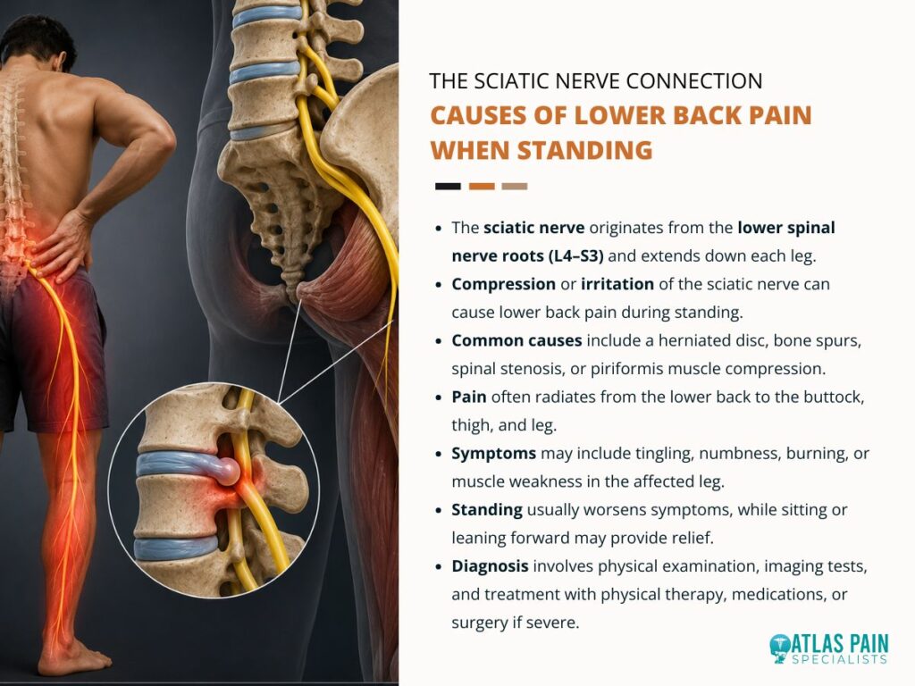
5. The Sciatic Nerve Connection
The sciatic nerve originates from the L4 through S3 nerve roots in the lower spine. This nerve travels through the pelvis and down the posterior aspect of each leg.
Compression or irritation of the sciatic nerve produces pain that radiates along its pathway. Standing can exacerbate this compression by narrowing the intervertebral foramina through which the nerve roots exit.
Mechanisms of Nerve Compression
A herniated disc, bone spur, or narrowed spinal canal can impinge on the sciatic nerve roots. The piriformis muscle in the buttock can also compress the nerve as it passes underneath or through the muscle.
The position of standing increases lumbar extension in some individuals. This extension reduces the space available for the nerve roots within the neural foramina.
Symptom Presentation
Sciatic pain presents as a sharp, shooting sensation that travels from the lower back into the buttock and leg. The pain may be accompanied by tingling, numbness, or a burning sensation in the affected leg.
Symptoms often worsen with standing and improve with sitting or lying down. Flexion of the spine, such as leaning forward, can reduce the nerve compression and provide relief.
Clinical Evaluation
The straight leg raise test helps identify sciatic nerve irritation. This test involves passive elevation of the straightened leg while the patient lies supine.
A positive test reproduces radiating pain down the leg and suggests nerve root involvement. Imaging studies confirm the anatomical source of compression and guide treatment decisions.
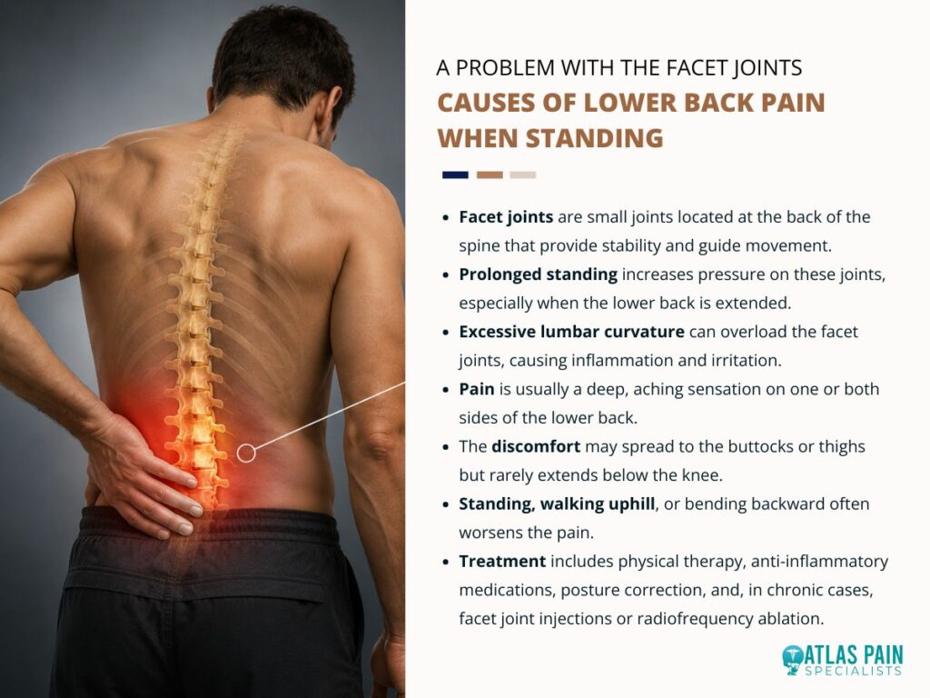
6. A Problem With the Facet Joints
Facet joints are paired structures located at the posterior aspect of each vertebral segment. These joints guide spinal motion and provide stability during flexion and extension movements.
Standing in an extended position compresses these joints against each other. This compression generates pain through mechanical stress and local inflammation.
Facet Joint Mechanics
Each facet joint consists of articular cartilage and a synovial lining. The joint capsules contain nerve endings that signal pain when stretched or compressed.
Excessive lumbar lordosis increases the load on the facet joints. This load can cause synovial impingement and capsular irritation over time.
Pain Characteristics
Facet joint pain presents as a deep ache localized to one or both sides of the lumbar spine. The pain does not typically radiate into the leg but may refer to the buttock or thigh.
Extension activities such as standing or walking uphill worsen the symptoms. Flexion of the spine, such as bending forward, provides relief by unloading the joints.
Diagnostic Procedures
Diagnostic facet joint blocks confirm the joint as the pain source. These injections contain local anesthetic placed directly into the joint capsule.
A positive response to the block indicates that the facet joint contributes to the patient's pain. Treatment includes physical therapy, anti-inflammatory medications, and radiofrequency ablation in chronic cases.
7. Spinal Stenosis and Narrowing Space
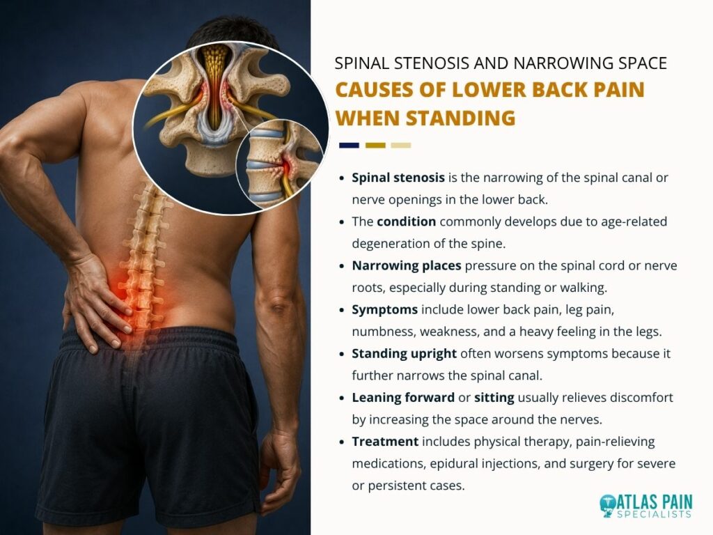
Spinal stenosis refers to a reduction in the available space within the spinal canal or neural foramina. This narrowing can compress the spinal cord or nerve roots and produce symptoms during standing.
The degenerative form of stenosis results from hypertrophic changes in the facet joints and ligamentum flavum. These structures thicken with age and encroach upon the neural elements.
Central Canal Stenosis
Central stenosis affects the main spinal canal and its contents. Standing with the spine in extension reduces the canal's diameter and worsens nerve compression.
Patients with central stenosis often experience bilateral leg symptoms. These symptoms include pain, weakness, and a heavy sensation in the lower extremities.
Lateral Recess Stenosis
Lateral recess stenosis involves narrowing of the side pockets of the spinal canal. This condition affects the exiting nerve roots as they traverse the lateral recess.
Symptoms present unilaterally and correspond to the specific nerve root affected. The L4 and L5 roots are most frequently involved.
Relief Through Flexion
Patients with stenosis frequently report relief when leaning forward over a shopping cart. This posture increases the spinal canal diameter by creating spinal flexion.
Sitting also reduces symptoms by unloading the spine and opening the canal space. These positional changes serve as important diagnostic clues.
8. Ankylosing Spondylitis and Inflammation
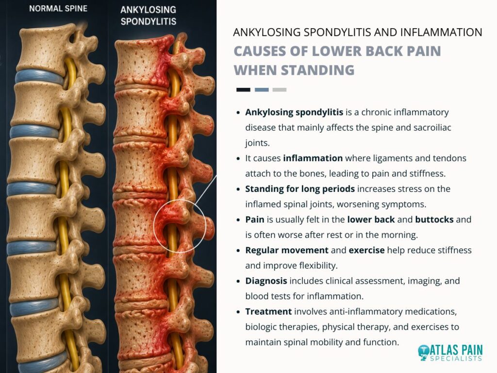
Ankylosing spondylitis is a chronic inflammatory condition that primarily affects the sacroiliac joints and the spine. This autoimmune disease causes enthesitis, which is inflammation at the sites where ligaments and tendons attach to bone.
Standing places axial load on inflamed spinal structures and exacerbates the pain. The inflammatory process leads to progressive stiffness and eventual bony fusion of the vertebral segments.
Disease Mechanism and Onset
The condition has a strong genetic association with the HLA-B27 antigen. This genetic marker appears in over ninety percent of affected individuals.
Inflammation begins at the sacroiliac joints and ascends the spine over years. New bone formation occurs at the margins of the vertebral bodies and produces syndesmophytes.
Pain Characteristics
Inflammatory back pain differs from mechanical back pain in several ways. The pain improves with physical activity and worsens with prolonged rest or stillness.
Morning stiffness lasts longer than thirty minutes after waking. Standing for extended periods triggers deep pain in the lower back and buttocks.
Diagnostic and Treatment Approach
Diagnosis relies on radiographic evidence of sacroiliitis and clinical criteria. Elevated inflammatory markers such as erythrocyte sedimentation rate support the diagnosis.
Treatment includes nonsteroidal anti-inflammatory drugs and biologic agents. Physical therapy maintains spinal mobility and delays the progression of fusion.
Conclusion
Lower back pain during standing arises from a diverse set of mechanical, degenerative, and inflammatory processes. Accurate diagnosis requires a thorough clinical history and physical examination to distinguish between these distinct etiologies.
Self-treatment without professional guidance carries the risk of worsening the underlying condition. A qualified healthcare provider can perform specific tests to isolate the pain source and recommend appropriate interventions.
Treatment strategies range from postural re-education and targeted strengthening to pharmacological management and surgical options. Patients who experience persistent or progressive symptoms should seek medical evaluation without delay.
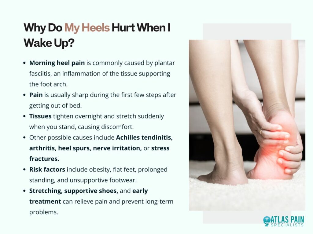
If you find yourself taking painful first steps out of bed every morning, the most common reason is inflammation of the tissues that support your heel, particularly a condition called plantar fasciitis.
Many people experience sharp, stabbing, or aching heel pain when they first stand up after sleeping. The discomfort often feels worst during those initial steps and gradually improves as they begin moving around.
While plantar fasciitis is the leading cause, several other conditions can also explain why do my heels hurt when I wake up, including Achilles tendon problems, arthritis, nerve irritation, and changes in foot structure.
Morning heel pain is more than an inconvenience. It can affect your ability to exercise, work comfortably, and stay active throughout the day. Why do my heels hurt when I wake up? Understanding what causes this symptom is the first step toward finding relief and preventing the problem from becoming chronic.
Understanding Morning Heel Pain
Morning heel pain usually occurs because tissues in the foot tighten during periods of rest and become stressed when weight is suddenly placed on them. During sleep, your feet remain relatively still for several hours. Tendons, ligaments, and connective tissues can shorten slightly while you rest. When you stand up and take your first steps, these tissues stretch abruptly, creating pain and stiffness.
This explains why many people notice a distinct pattern. The pain is intense when getting out of bed but decreases after several minutes of walking. Unfortunately, the discomfort often returns after sitting for long periods during the day.
The heel is designed to absorb significant force. Every step places pressure on the heel bone, surrounding tissues, and supporting structures. When inflammation develops, even normal walking can become painful.
While occasional soreness may result from overuse or an unusually active day, persistent morning heel pain often signals an underlying condition that deserves attention.
Plantar Fasciitis: The Most Common Cause of Morning Heel Pain
Plantar fasciitis is the leading reason people ask, "why do my heels hurt when I wake up?" The plantar fascia is a thick band of connective tissue that runs from the heel bone to the toes. It supports the arch of the foot and helps absorb shock during walking and running.
When this tissue becomes irritated or develops tiny tears, inflammation occurs.
Overnight, the plantar fascia tightens slightly. The first steps in the morning stretch the inflamed tissue, creating the characteristic sharp pain many people experience.
Common Symptoms of Plantar Fasciitis
- Sharp pain under the heel
- Pain that is worst with the first steps of the day
- Discomfort after long periods of sitting
- Heel tenderness when pressed
- Pain that improves somewhat with movement
Many people describe the sensation as stepping on a nail or experiencing a sudden stabbing pain beneath the heel.
Certain factors increase the risk of plantar fasciitis:
| Risk Factor | How It Contributes |
| Obesity | Increases pressure on the plantar fascia |
| Flat feet | Places additional strain on foot structures |
| High arches | Reduces shock absorption |
| Running or jumping sports | Repetitive stress on the heel |
| Standing for long hours | Constant strain on the fascia |
| Poor footwear | Insufficient support and cushioning |
Without treatment, plantar fasciitis can persist for months and sometimes become a long-term problem.
Other Conditions That Can Cause Heel Pain in the Morning
Although plantar fasciitis is common, several other conditions can cause similar morning heel pain. Accurate diagnosis matters because treatment varies depending on the underlying cause.
Achilles Tendinitis
The Achilles tendon connects the calf muscles to the heel bone. Repetitive stress can cause inflammation and stiffness, particularly in runners and active individuals.
People with Achilles tendinitis often notice:
- Pain at the back of the heel
- Morning stiffness
- Tenderness along the tendon
- Increased pain after activity
The discomfort typically improves as the tendon warms up but may worsen later in the day.
Heel Spurs
Heel spurs are bony growths that develop on the underside of the heel bone. While many heel spurs cause no symptoms, some contribute to pain when combined with plantar fasciitis.
The spur itself is not always the source of discomfort. Instead, the associated inflammation around the area often causes symptoms.
Arthritis
Several forms of arthritis can affect the feet and heels.
Conditions that may contribute include:
- Osteoarthritis
- Rheumatoid arthritis
- Psoriatic arthritis
- Reactive arthritis
Inflammation within joints and surrounding tissues can create morning stiffness and pain that gradually improves with movement.
Nerve Entrapment
Compressed nerves around the ankle or heel may cause pain, tingling, burning, or numbness.
Unlike plantar fasciitis, nerve-related pain often feels electrical or radiates into other parts of the foot. Symptoms may occur throughout the day rather than only during the first morning steps.
Stress Fractures
Small cracks in the heel bone can develop due to repetitive impact.
Athletes, military personnel, and individuals who suddenly increase activity levels may be at higher risk. Pain from a stress fracture typically worsens with activity and may persist even during rest.
Factors That Increase Your Risk of Morning Heel Pain
Certain lifestyle and health factors make morning heel pain more likely to develop. Understanding these risk factors can help identify the root cause and prevent future problems.
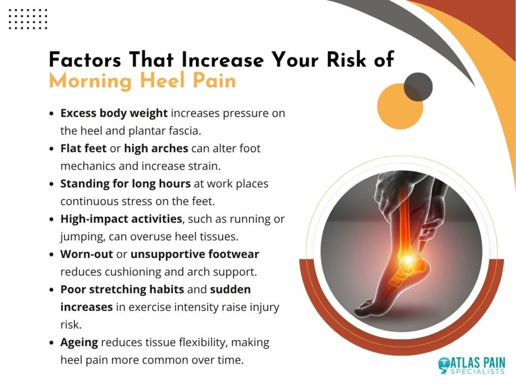
Weight and Foot Mechanics
Excess body weight increases the load placed on the feet every day. Even modest weight gain can significantly increase pressure on the plantar fascia and heel structures. Over time, this additional stress contributes to inflammation and tissue breakdown.
Foot structure also matters. Both flat feet and high arches can alter weight distribution and create abnormal stress patterns.
Occupation and Daily Activities
People who spend long hours standing often develop heel pain more frequently than those who sit for most of the day.
High-risk occupations include:
- Teachers
- Nurses
- Factory workers
- Retail employees
- Construction workers
Repeated loading without adequate recovery can gradually damage supporting tissues.
Exercise Habits
Physical activity is beneficial, but sudden increases in intensity can strain the heel.
Common exercise-related contributors include:
- Running on hard surfaces
- Excessive jumping
- Poor training progression
- Worn-out athletic shoes
- Inadequate stretching
Many athletes first notice heel pain after increasing mileage or changing training routines.
Aging
As people age, the tissues supporting the foot naturally lose some elasticity and shock-absorbing capacity.
This makes inflammation and overuse injuries more likely. Morning heel pain becomes increasingly common after age 40, although younger individuals can certainly develop it as well.
How Doctors Diagnose the Cause of Heel Pain
A proper diagnosis helps determine why your heels hurt when you wake up and guides effective treatment. Diagnosis usually begins with a detailed medical history and physical examination.
A healthcare provider may ask:
- When the pain started
- Where the pain is located
- What activities worsen symptoms
- Whether pain improves with movement
- If numbness or tingling occurs
The location of pain often provides important clues. Pain underneath the heel frequently suggests plantar fasciitis, while pain at the back of the heel may indicate Achilles tendon problems.
In some cases, imaging studies are recommended.
| Diagnostic Tool | Purpose |
| X-rays | Identify fractures or heel spurs |
| Ultrasound | Evaluate soft tissue inflammation |
| MRI | Assess tendons, fascia, and bone injuries |
| Blood tests | Investigate inflammatory arthritis |
Most cases of plantar fasciitis can be diagnosed without advanced imaging, but additional testing may be needed when symptoms are unusual or persistent.
Effective Treatments for Morning Heel Pain
The good news is that most causes of morning heel pain improve with conservative treatment. Treatment focuses on reducing inflammation, improving flexibility, and addressing contributing factors.
Stretching Exercises
Stretching is often one of the most effective interventions.
Regular stretching can help:
- Lengthen the plantar fascia
- Improve calf flexibility
- Reduce morning stiffness
- Decrease tension on the heel
Many healthcare providers recommend stretching before getting out of bed to minimize pain during those first steps.
Supportive Footwear
Shoes play a major role in recovery.
Supportive footwear should provide:
- Arch support
- Adequate cushioning
- Shock absorption
- Stability
Walking barefoot on hard surfaces often worsens symptoms and should be limited during recovery.
Ice Therapy
Applying ice to the heel can help reduce inflammation. Many people find relief by rolling their foot over a frozen water bottle for 10 to 15 minutes. This combines icing with gentle stretching.
Orthotics and Inserts
Custom or over-the-counter orthotics can improve foot alignment and reduce strain on the plantar fascia. These devices help distribute pressure more evenly and provide additional support during daily activities.
Physical Therapy
Physical therapy may be recommended when symptoms persist.
Treatment often includes:
- Stretching programs
- Strengthening exercises
- Manual therapy
- Gait assessment
- Activity modification
Many patients experience significant improvement after following a structured rehabilitation plan.
When Morning Heel Pain Requires Medical Attention
Persistent heel pain should not be ignored, especially if symptoms continue despite home treatment. While many cases improve with conservative care, certain warning signs warrant prompt evaluation.
Seek medical attention if you experience:
- Severe pain that limits walking
- Sudden swelling
- Numbness or weakness
- Fever or redness
- Pain following a significant injury
- Symptoms lasting several weeks without improvement
Early treatment often prevents chronic problems and helps people return to normal activities more quickly. Ignoring ongoing pain can lead to compensatory walking patterns that create additional issues in the knees, hips, and lower back.
Preventing Future Heel Pain
Preventing recurring heel pain involves reducing stress on the foot while maintaining flexibility and strength. Simple daily habits can make a substantial difference.
Consider the following preventive strategies:
- Maintain a healthy weight
- Wear supportive footwear
- Replace worn athletic shoes regularly
- Stretch calves and feet daily
- Increase exercise intensity gradually
- Avoid prolonged barefoot walking on hard surfaces
Consistency matters more than intensity. Small preventive measures performed regularly often provide the greatest long-term benefit.
Conclusion
If you're wondering why do my heels hurt when I wake up, the answer is most often related to plantar fasciitis, although Achilles tendon issues, arthritis, nerve problems, and other conditions can also contribute.
Morning heel pain occurs because tissues tighten during rest and become stressed when you first place weight on your feet. While the discomfort can be frustrating, most cases respond well to stretching, supportive footwear, activity modifications, and other conservative treatments.
The key is addressing symptoms early rather than waiting for them to worsen. By identifying the underlying cause and following an appropriate treatment plan, most people can reduce pain, improve mobility, and start their mornings without those painful first steps.
Lower back pain when walking but not sitting is often caused by conditions that place stress on the spine, joints, muscles, or nerves during movement and weight-bearing activities. While walking is generally beneficial for spinal health, some people experience discomfort that appears only when they are on their feet.
The pain may start as a mild ache, stiffness, or pressure in the lower back and gradually worsen with continued movement. Sitting frequently provides relief because it changes spinal positioning and reduces stress on certain structures.
Why does my lower back hurt when walking but not sitting? Understanding the causes of lower back pain when walking can help identify underlying issues, recognize warning signs, and determine the most effective treatment options for lasting relief.
How Walking Affects the Lower Back
Walking places repeated stress on the structures that support your spine, making underlying problems more noticeable.The lower back is designed to support body weight while allowing flexibility and movement. Every step you take creates forces that travel through the feet, legs, pelvis, and spine. Normally, muscles, discs, ligaments, and joints work together to absorb these forces efficiently.
When one of these structures becomes irritated or damaged, walking may trigger pain. Unlike sitting, which often unloads certain spinal tissues, walking requires continuous muscle engagement and weight-bearing.
This increased demand can expose weaknesses or injuries that remain unnoticed while resting.The pattern of pain can provide important clues.
Pain that worsens steadily with walking but improves quickly when sitting often points toward specific spinal conditions, while pain that occurs only after long distances may indicate muscular fatigue or biomechanical issues.
Understanding these movement-related changes helps explain why symptoms appear during activity but disappear when resting.
Common Causes of Lower Back Pain When Walking
Several conditions can cause lower back pain when walking, but some are far more common than others.

Lumbar Spinal Stenosis
Lumbar spinal stenosis is one of the most frequent causes of lower back pain when walking, particularly in older adults.This condition occurs when the spaces within the spine narrow, placing pressure on nerves.
As you walk, the spine naturally extends slightly, which can further narrow these spaces and increase nerve compression.
People with spinal stenosis often notice:
- Back pain during walking
- Leg pain or heaviness
- Numbness or tingling
- Symptoms that improve when sitting
- Relief when leaning forward
Many individuals find they can walk farther when pushing a shopping cart because the forward-leaning position opens the spinal canal and reduces pressure on nerves.
Degenerative Disc Disease
The discs between vertebrae act as shock absorbers. Over time, these discs naturally lose water content and become less effective.When degeneration occurs, walking can place increased stress on nearby joints and nerves.
This may create pain that worsens with prolonged standing or walking but improves with sitting. Degenerative disc disease often develops gradually. Some people experience occasional discomfort, while others develop chronic pain that limits daily activities.
Facet Joint Arthritis
Facet joints connect the vertebrae and guide spinal movement. Like other joints in the body, they can develop arthritis over time. Walking increases movement through these joints, which may trigger inflammation and discomfort.
Sitting often provides relief because it reduces joint loading.
Facet joint pain is frequently described as:
- Deep aching pain
- Localized lower back discomfort
- Stiffness after activity
- Pain that worsens when standing upright
The discomfort may become more noticeable after long walks or periods of prolonged standing.
Sacroiliac Joint Dysfunction
The sacroiliac joints connect the spine to the pelvis. When these joints become inflamed or unstable, walking can trigger significant discomfort.
Each step transfers force through the pelvis, causing repetitive stress on the sacroiliac joints. Sitting reduces this movement, often resulting in symptom relief.
People with sacroiliac dysfunction frequently report pain in the lower back, buttocks, or hips. Sometimes the discomfort extends into the upper leg, making diagnosis more challenging.
Muscular Causes of Lower Back Pain When Walking
Not all lower back pain originates from the spine itself; muscles often play a major role. Muscles surrounding the spine provide stability and support during movement.
Weakness, tightness, or fatigue in these muscles can lead to pain that becomes noticeable during walking. When muscles are unable to adequately support the spine, other structures may compensate, increasing stress throughout the lower back.
Common muscular contributors include:
| Muscular Issue | How It Causes Pain |
| Muscle strain | Inflamed fibers become painful during movement |
| Core weakness | Reduced spinal stability during walking |
| Tight hip flexors | Increased stress on the lower back |
| Glute weakness | Poor pelvic support |
| Poor posture | Uneven force distribution |
Many people spend long hours sitting during the day, which weakens core muscles and tightens hip structures. When they begin walking, these imbalances place extra strain on the lower back.
The good news is that muscular causes often respond well to exercise, stretching, and strengthening programs.
Nerve-Related Conditions That Cause Pain While Walking
Nerve irritation can produce lower back pain that becomes significantly worse during movement.
The nerves that exit the lower spine control sensation and movement throughout the lower body. When these nerves become compressed or irritated, walking may aggravate symptoms.
Sciatica
Sciatica occurs when the sciatic nerve becomes irritated or compressed.
Although many people think of sciatica as leg pain, it often begins in the lower back. Walking can increase nerve tension and trigger discomfort that radiates through the buttocks and legs.
Symptoms may include:
- Sharp lower back pain
- Burning sensations
- Tingling
- Numbness
- Pain extending below the knee
Some individuals can walk only short distances before symptoms force them to stop and rest.
Herniated Disc
A herniated disc develops when the soft inner portion of a spinal disc pushes outward and irritates nearby nerves.
Depending on the location of the herniation, walking may increase nerve pressure and worsen symptoms. Sitting sometimes reduces irritation by changing spinal mechanics and relieving stress on affected tissues.
Not every herniated disc causes pain, but when nerve compression occurs, walking often becomes uncomfortable.
Why Sitting Often Relieves the Pain
The fact that sitting reduces discomfort provides important clues about the underlying cause.
When you sit, several changes occur within the spine:
- Body weight is redistributed
- Certain spinal joints experience less pressure
- Nerve compression may decrease
- Muscles can relax
- Walking-related forces are removed
For conditions such as spinal stenosis, sitting often opens the spaces surrounding spinal nerves, providing immediate relief. For muscular conditions, sitting reduces workload and allows fatigued tissues to recover temporarily.
This relationship between walking and sitting helps healthcare providers narrow down possible diagnoses during evaluation.
Signs That Your Lower Back Pain May Be Serious
While many causes of lower back pain are manageable, certain symptoms should never be ignored.
Most episodes of lower back pain when walking improve with conservative treatment. However, some warning signs indicate the need for prompt medical attention.
Seek medical evaluation if you experience:
- Progressive leg weakness
- Loss of bladder control
- Loss of bowel control
- Severe numbness in the groin area
- Unexplained weight loss
- Fever accompanying back pain
- Persistent nighttime pain
- Recent significant trauma
These symptoms could indicate serious neurological or systemic conditions requiring immediate care. Even without emergency symptoms, pain lasting several weeks should be evaluated by a healthcare professional.
Diagnosing the Cause of Lower Back Pain When Walking
Accurate diagnosis is essential because treatment depends on identifying the true source of the pain.
Healthcare providers typically begin with a detailed medical history and physical examination. They often ask specific questions about:
- When the pain started
- How far you can walk before symptoms begin
- Whether sitting relieves pain
- Any leg symptoms
- Previous injuries
The pattern of symptoms often provides valuable diagnostic clues.
Additional testing may include:
| Diagnostic Test | Purpose |
| X-rays | Evaluate bones and arthritis |
| MRI | Assess discs, nerves, and soft tissues |
| CT scan | Examine spinal structures |
| Nerve studies | Evaluate nerve function |
| Diagnostic injections | Identify pain sources |
Imaging is not always necessary immediately. Many cases improve with conservative management before advanced testing becomes necessary.
Treatment Options for Lower Back Pain When Walking
Treatment focuses on reducing pain, improving function, and addressing the underlying cause. The most effective treatment plan depends on the diagnosis. However, several approaches commonly help people experiencing lower back pain when walking.

Physical Therapy
Physical therapy is a common treatment for lower back pain when walking because it helps address muscle weakness, poor posture, and movement issues that contribute to discomfort.
Through targeted exercises and movement training, physical therapy strengthens supporting muscles, improves flexibility and balance, and reduces stress on the spine, helping improve walking tolerance and overall mobility. .
Activity Modification
Temporarily adjusting activity levels may allow irritated tissues to heal. This does not necessarily mean complete rest. In fact, prolonged inactivity often worsens back pain.
Instead, activity modification involves finding a comfortable level of movement while avoiding excessive strain. Many people benefit from shorter walks performed more frequently throughout the day.
Medications
Depending on the cause, medications may help reduce symptoms.
Common options include:
- Anti-inflammatory medications
- Muscle relaxants
- Nerve pain medications
- Topical pain relievers
Medication should be viewed as one component of treatment rather than a complete solution.
Injections
For some conditions, injections provide both diagnostic information and symptom relief.
Examples include:
- Epidural steroid injections
- Facet joint injections
- Sacroiliac joint injections
These treatments can reduce inflammation and improve mobility, especially when combined with rehabilitation.
Surgical Treatment
Surgery is usually considered only when conservative treatments fail or when significant nerve compression exists.Procedures vary depending on the condition but may involve removing pressure from nerves or stabilizing spinal structures.
Fortunately, many people improve without needing surgery.
Ways to Prevent Lower Back Pain When Walking
Simple lifestyle habits can significantly reduce the risk of developing recurring lower back pain.
Prevention focuses on maintaining spinal health and reducing unnecessary stress on supporting structures.
Helpful strategies include:
- Maintaining a healthy body weight
- Exercising regularly
- Strengthening core muscles
- Stretching hips and hamstrings
- Wearing supportive footwear
- Practicing good posture
- Avoiding prolonged sitting
These habits improve spinal stability and reduce the likelihood of pain developing during activity. Consistency is more important than intensity. Small daily efforts often produce better long-term results than occasional aggressive exercise programs.
Conclusion
Lower back pain when walking but not sitting is often a sign that certain structures in the spine, joints, muscles, or nerves are being stressed during movement and weight-bearing activities. Conditions such as lumbar spinal stenosis, degenerative disc disease, facet joint arthritis, sacroiliac joint dysfunction, muscular imbalances, and nerve compression can all contribute to this unique pattern of pain.
While sitting frequently provides relief by reducing pressure on these structures, the recurring discomfort during walking should not be ignored.The good news is that most cases respond well to appropriate treatment, especially when the underlying cause is identified early.
Physical therapy, targeted exercise, posture improvements, activity modifications, and other conservative treatments often help restore mobility and reduce pain significantly. If symptoms persist, worsen, or are accompanied by numbness, weakness, or other neurological signs, professional evaluation is important.
Understanding why the pain occurs is the first step toward finding effective treatment and returning to comfortable, confident movement.
Lower back pain when walking but not sitting is often caused by conditions that place stress on the spine, joints, muscles, or nerves during movement and weight-bearing activities. While walking is generally beneficial for spinal health, some people experience discomfort that appears only when they are on their feet.
The pain may start as a mild ache, stiffness, or pressure in the lower back and gradually worsen with continued movement. Sitting frequently provides relief because it changes spinal positioning and reduces stress on certain structures.
Understanding the causes of lower back pain when walking can help identify underlying issues, recognize warning signs, and determine the most effective treatment options for lasting relief.
How Walking Affects the Lower Back
Walking places repeated stress on the structures that support your spine, making underlying problems more noticeable.The lower back is designed to support body weight while allowing flexibility and movement. Every step you take creates forces that travel through the feet, legs, pelvis, and spine. Normally, muscles, discs, ligaments, and joints work together to absorb these forces efficiently.
When one of these structures becomes irritated or damaged, walking may trigger pain. Unlike sitting, which often unloads certain spinal tissues, walking requires continuous muscle engagement and weight-bearing.
This increased demand can expose weaknesses or injuries that remain unnoticed while resting.The pattern of pain can provide important clues.
Pain that worsens steadily with walking but improves quickly when sitting often points toward specific spinal conditions, while pain that occurs only after long distances may indicate muscular fatigue or biomechanical issues.
Understanding these movement-related changes helps explain why symptoms appear during activity but disappear when resting.
Common Causes of Lower Back Pain When Walking
Several conditions can cause lower back pain when walking, but some are far more common than others.
Lumbar Spinal Stenosis
Lumbar spinal stenosis is one of the most frequent causes of lower back pain when walking, particularly in older adults.This condition occurs when the spaces within the spine narrow, placing pressure on nerves.
As you walk, the spine naturally extends slightly, which can further narrow these spaces and increase nerve compression.
People with spinal stenosis often notice:
- Back pain during walking
- Leg pain or heaviness
- Numbness or tingling
- Symptoms that improve when sitting
- Relief when leaning forward
Many individuals find they can walk farther when pushing a shopping cart because the forward-leaning position opens the spinal canal and reduces pressure on nerves.
Degenerative Disc Disease
The discs between vertebrae act as shock absorbers. Over time, these discs naturally lose water content and become less effective.When degeneration occurs, walking can place increased stress on nearby joints and nerves.
This may create pain that worsens with prolonged standing or walking but improves with sitting. Degenerative disc disease often develops gradually. Some people experience occasional discomfort, while others develop chronic pain that limits daily activities.
Facet Joint Arthritis
Facet joints connect the vertebrae and guide spinal movement. Like other joints in the body, they can develop arthritis over time. Walking increases movement through these joints, which may trigger inflammation and discomfort.
Sitting often provides relief because it reduces joint loading.
Facet joint pain is frequently described as:
- Deep aching pain
- Localized lower back discomfort
- Stiffness after activity
- Pain that worsens when standing upright
The discomfort may become more noticeable after long walks or periods of prolonged standing.
Sacroiliac Joint Dysfunction
The sacroiliac joints connect the spine to the pelvis. When these joints become inflamed or unstable, walking can trigger significant discomfort.
Each step transfers force through the pelvis, causing repetitive stress on the sacroiliac joints. Sitting reduces this movement, often resulting in symptom relief.
People with sacroiliac dysfunction frequently report pain in the lower back, buttocks, or hips. Sometimes the discomfort extends into the upper leg, making diagnosis more challenging.
Muscular Causes of Lower Back Pain When Walking
Not all lower back pain originates from the spine itself; muscles often play a major role. Muscles surrounding the spine provide stability and support during movement.
Weakness, tightness, or fatigue in these muscles can lead to pain that becomes noticeable during walking. When muscles are unable to adequately support the spine, other structures may compensate, increasing stress throughout the lower back.
Common muscular contributors include:
| Muscular Issue | How It Causes Pain |
| Muscle strain | Inflamed fibers become painful during movement |
| Core weakness | Reduced spinal stability during walking |
| Tight hip flexors | Increased stress on the lower back |
| Glute weakness | Poor pelvic support |
| Poor posture | Uneven force distribution |
Many people spend long hours sitting during the day, which weakens core muscles and tightens hip structures. When they begin walking, these imbalances place extra strain on the lower back.
The good news is that muscular causes often respond well to exercise, stretching, and strengthening programs.
Nerve-Related Conditions That Cause Pain While Walking
Nerve irritation can produce lower back pain that becomes significantly worse during movement.
The nerves that exit the lower spine control sensation and movement throughout the lower body. When these nerves become compressed or irritated, walking may aggravate symptoms.
Sciatica
Sciatica occurs when the sciatic nerve becomes irritated or compressed.
Although many people think of sciatica as leg pain, it often begins in the lower back. Walking can increase nerve tension and trigger discomfort that radiates through the buttocks and legs.
Symptoms may include:
- Sharp lower back pain
- Burning sensations
- Tingling
- Numbness
- Pain extending below the knee
Some individuals can walk only short distances before symptoms force them to stop and rest.
Herniated Disc
A herniated disc develops when the soft inner portion of a spinal disc pushes outward and irritates nearby nerves.
Depending on the location of the herniation, walking may increase nerve pressure and worsen symptoms. Sitting sometimes reduces irritation by changing spinal mechanics and relieving stress on affected tissues.
Not every herniated disc causes pain, but when nerve compression occurs, walking often becomes uncomfortable.
Why Sitting Often Relieves the Pain
The fact that sitting reduces discomfort provides important clues about the underlying cause.
When you sit, several changes occur within the spine:
- Body weight is redistributed
- Certain spinal joints experience less pressure
- Nerve compression may decrease
- Muscles can relax
- Walking-related forces are removed
For conditions such as spinal stenosis, sitting often opens the spaces surrounding spinal nerves, providing immediate relief. For muscular conditions, sitting reduces workload and allows fatigued tissues to recover temporarily.
This relationship between walking and sitting helps healthcare providers narrow down possible diagnoses during evaluation.
Signs That Your Lower Back Pain May Be Serious
While many causes of lower back pain are manageable, certain symptoms should never be ignored.
Most episodes of lower back pain when walking improve with conservative treatment. However, some warning signs indicate the need for prompt medical attention.
Seek medical evaluation if you experience:
- Progressive leg weakness
- Loss of bladder control
- Loss of bowel control
- Severe numbness in the groin area
- Unexplained weight loss
- Fever accompanying back pain
- Persistent nighttime pain
- Recent significant trauma
These symptoms could indicate serious neurological or systemic conditions requiring immediate care. Even without emergency symptoms, pain lasting several weeks should be evaluated by a healthcare professional.
Diagnosing the Cause of Lower Back Pain When Walking
Accurate diagnosis is essential because treatment depends on identifying the true source of the pain.
Healthcare providers typically begin with a detailed medical history and physical examination. They often ask specific questions about:
- When the pain started
- How far you can walk before symptoms begin
- Whether sitting relieves pain
- Any leg symptoms
- Previous injuries
The pattern of symptoms often provides valuable diagnostic clues.
Additional testing may include:
| Diagnostic Test | Purpose |
| X-rays | Evaluate bones and arthritis |
| MRI | Assess discs, nerves, and soft tissues |
| CT scan | Examine spinal structures |
| Nerve studies | Evaluate nerve function |
| Diagnostic injections | Identify pain sources |
Imaging is not always necessary immediately. Many cases improve with conservative management before advanced testing becomes necessary.
Treatment Options for Lower Back Pain When Walking
Treatment focuses on reducing pain, improving function, and addressing the underlying cause. The most effective treatment plan depends on the diagnosis. However, several approaches commonly help people experiencing lower back pain when walking.
Physical Therapy
Physical therapy is a common treatment for lower back pain when walking because it helps address muscle weakness, poor posture, and movement issues that contribute to discomfort.
Through targeted exercises and movement training, physical therapy strengthens supporting muscles, improves flexibility and balance, and reduces stress on the spine, helping improve walking tolerance and overall mobility. .
Activity Modification
Temporarily adjusting activity levels may allow irritated tissues to heal. This does not necessarily mean complete rest. In fact, prolonged inactivity often worsens back pain.
Instead, activity modification involves finding a comfortable level of movement while avoiding excessive strain. Many people benefit from shorter walks performed more frequently throughout the day.
Medications
Depending on the cause, medications may help reduce symptoms.
Common options include:
- Anti-inflammatory medications
- Muscle relaxants
- Nerve pain medications
- Topical pain relievers
Medication should be viewed as one component of treatment rather than a complete solution.
Injections
For some conditions, injections provide both diagnostic information and symptom relief.
Examples include:
- Epidural steroid injections
- Facet joint injections
- Sacroiliac joint injections
These treatments can reduce inflammation and improve mobility, especially when combined with rehabilitation.
Surgical Treatment
Surgery is usually considered only when conservative treatments fail or when significant nerve compression exists.Procedures vary depending on the condition but may involve removing pressure from nerves or stabilizing spinal structures.
Fortunately, many people improve without needing surgery.
Ways to Prevent Lower Back Pain When Walking
Simple lifestyle habits can significantly reduce the risk of developing recurring lower back pain.
Prevention focuses on maintaining spinal health and reducing unnecessary stress on supporting structures.
Helpful strategies include:
- Maintaining a healthy body weight
- Exercising regularly
- Strengthening core muscles
- Stretching hips and hamstrings
- Wearing supportive footwear
- Practicing good posture
- Avoiding prolonged sitting
These habits improve spinal stability and reduce the likelihood of pain developing during activity. Consistency is more important than intensity. Small daily efforts often produce better long-term results than occasional aggressive exercise programs.
Conclusion
Lower back pain when walking but not sitting is often a sign that certain structures in the spine, joints, muscles, or nerves are being stressed during movement and weight-bearing activities. Conditions such as lumbar spinal stenosis, degenerative disc disease, facet joint arthritis, sacroiliac joint dysfunction, muscular imbalances, and nerve compression can all contribute to this unique pattern of pain.
While sitting frequently provides relief by reducing pressure on these structures, the recurring discomfort during walking should not be ignored.The good news is that most cases respond well to appropriate treatment, especially when the underlying cause is identified early.
Physical therapy, targeted exercise, posture improvements, activity modifications, and other conservative treatments often help restore mobility and reduce pain significantly. If symptoms persist, worsen, or are accompanied by numbness, weakness, or other neurological signs, professional evaluation is important.
Understanding why the pain occurs is the first step toward finding effective treatment and returning to comfortable, confident movement.
Osteoporosis is often called a “silent disease” because it usually does not cause pain until a fracture or complication occurs. While osteoporosis itself does not directly cause leg pain, weakened bones can lead to fractures in the hip, pelvis, or spine that may trigger pain in the legs, groin, or lower back.
Changes in posture, balance, and movement can also strain muscles and nerves, causing discomfort. In some cases, leg pain may result from related conditions such as arthritis or sciatica.
Is osteoporosis painful in the legs? Because fragile bones break more easily, persistent or unexplained leg pain should never be ignored, especially after a fall or minor injury.
Understanding Osteoporosis and Pain
Osteoporosis is a bone disease that weakens bones by reducing bone density and strength, making fractures more likely. Healthy bones are strong enough to support body weight and absorb normal stress. With osteoporosis, bones become thinner, more fragile, and more vulnerable to breaks from falls, small impacts, or even ordinary movements in severe cases.

Why Osteoporosis Is Often Silent
In the early stages, osteoporosis usually does not cause obvious symptoms. A person may feel completely normal while bone density gradually declines. This is why many people do not realize they have osteoporosis until they experience a fracture.
There are often no symptoms in the early stages of bone loss, but once bones weaken, signs may include back pain from a broken or collapsed spinal bone, height loss, stooped posture, or a bone breaking more easily than expected.
This silent progression can make osteoporosis confusing. People may expect weak bones to ache constantly, but bone loss itself generally does not create pain signals until bone structure is damaged.
When Pain Begins
Pain usually begins when osteoporosis causes a fracture or contributes to another physical problem. The National Institute of Arthritis and Musculoskeletal and Skin Diseases explains that osteoporosis is often called a silent disease because there are typically no symptoms until a bone is broken.
Vertebral fractures may cause severe back pain, height loss, or a stooped posture.This means leg pain in someone with osteoporosis should be evaluated carefully.
It may be related to a fracture in the hip, pelvis, thigh bone, or spine, but it may also come from muscles, joints, nerves, or circulation.
Is Osteoporosis Painful in the Legs?
Osteoporosis can be painful in the legs when it causes or contributes to fractures, but uncomplicated osteoporosis usually does not directly cause leg pain. This distinction matters because treating osteoporosis-related leg pain depends on identifying the actual source of the discomfort.
Direct vs Indirect Leg Pain
Direct osteoporosis-related leg pain usually happens when a weakened bone in or near the leg breaks. This may involve the hip, pelvis, femur, knee area, or ankle.
The pain may appear suddenly after a fall or develop gradually if there is a stress fracture. Indirect leg pain can occur when osteoporosis affects posture, balance, mobility, or spinal alignment.
For example, spinal compression fractures may change posture and walking mechanics, placing extra strain on the hips, thighs, knees, or calves. A compressed spinal bone may also irritate nearby nerves, leading to pain that travels into the legs.
Why Leg Pain Should Be Taken Seriously
Leg pain should be taken seriously when it is new, severe, unexplained, or linked to difficulty walking. In people with osteoporosis, even a minor fall can cause a fracture. Sometimes the injury may not seem dramatic at first, but pain can worsen with weight-bearing.
Osteoporosis makes bones more likely to break, and commonly affected fracture sites include the hip, spine, and wrist, although other bones can also break.
Common Osteoporosis-Related Causes of Leg Pain
Leg pain in people with osteoporosis is most often related to fractures, posture changes, or conditions that develop because the body is compensating for weakened bones. Understanding these patterns can help you decide when symptoms may need medical attention.
Hip Fractures
A hip fracture is one of the most serious osteoporosis-related injuries. It may happen after a fall, but in severe osteoporosis, even a small twist or awkward step can sometimes cause damage. Hip fracture pain may be felt in the hip, groin, thigh, or sometimes the knee.
The pain is usually worse when standing, walking, or trying to move the leg. Some people cannot bear weight on the affected side. The leg may also appear shortened or turned outward in more obvious fractures.
Hip fractures require urgent medical evaluation because delaying care can lead to complications and longer recovery.
Pelvic Fractures
Pelvic fractures can also cause pain that feels like it is coming from the leg. The pain may appear in the groin, buttock, lower back, hip, or upper thigh. It may worsen when walking, climbing stairs, or standing for long periods.
These fractures may be difficult to recognize because pain can be vague at first. Some people assume they have a muscle strain when the real issue is a small fracture in a weakened pelvic bone.
Femur or Thigh Bone Stress Fractures
The femur is the large bone in the thigh. Osteoporosis can increase the risk of stress fractures or more serious breaks in this area. Pain may develop gradually in the thigh and become worse with walking or standing.
Persistent thigh pain should be evaluated, especially if it occurs without a clear injury or worsens over time. This is particularly important for people already diagnosed with low bone density.
Spinal Compression Fractures
Spinal compression fractures usually cause back pain, but they can indirectly affect the legs. When vertebrae collapse or compress, posture and walking patterns may change. In some cases, nerve irritation may contribute to pain, weakness, tingling, or heaviness in the legs.
The Bone Health and Osteoporosis Foundation explains that spinal fractures can cause pain, height loss, and a stooped or hunched posture.
Comparing Leg Pain Causes in People With Osteoporosis
Not all leg pain in a person with osteoporosis is caused by osteoporosis. The table below shows common possibilities and how symptoms may differ.
| Possible Cause | Where Pain Is Felt | Common Pattern | Why It Matters |
| Hip fracture | Hip, groin, thigh, knee | Sudden pain, worse with weight-bearing | Needs urgent evaluation |
| Pelvic fracture | Groin, buttock, hip, upper thigh | Pain with walking or standing | Can be missed early |
| Femur stress fracture | Thigh or upper leg | Gradual pain that worsens with activity | May worsen if untreated |
| Spinal compression fracture | Back, sometimes legs | Back pain, posture change, possible nerve symptoms | May affect walking |
| Arthritis | Hip, knee, ankle | Aching stiffness, worse with use | Common alongside osteoporosis |
| Sciatica | Lower back, buttock, leg | Burning or shooting pain down one leg | Nerve-related, not bone density itself |
| Muscle strain | Thigh, calf, hip area | Soreness after activity | Usually improves with rest |
This comparison helps show why diagnosis matters. The same person can have osteoporosis and leg pain from arthritis, nerve irritation, or muscle strain at the same time.
Symptoms That Suggest Osteoporosis May Be Involved
Osteoporosis-related pain is most concerning when it suggests a fracture or structural change. Because osteoporosis may not hurt until a bone breaks, symptoms often appear suddenly or after a minor event.
Pain After a Fall or Minor Injury
Any new leg, hip, groin, or thigh pain after a fall should be taken seriously in someone with osteoporosis. Even if the fall seemed mild, weakened bones may fracture more easily than healthy bones.
Pain that worsens when standing, walking, or shifting weight may indicate a fracture. Rest may reduce pain temporarily, but the discomfort usually returns when the affected bone is stressed.
Sudden Difficulty Walking
Difficulty walking, limping, or inability to bear weight can be a warning sign. This is especially important if pain is located around the hip, pelvis, or upper thigh.
Some fractures are obvious immediately. Others cause more subtle pain that becomes worse over several hours or days.
Height Loss or Stooped Posture
Height loss and stooped posture do not directly cause leg pain, but they may indicate spinal fractures. Spinal changes can affect balance, walking pattern, and muscle strain in the hips and legs.
The Clinician’s Guide to Prevention and Treatment of Osteoporosis notes that repeat bone density testing and vertebral imaging may be considered when patients show signs of vertebral fracture, such as height loss or back pain.
What Leg Pain From a Fracture May Feel Like
Leg pain related to an osteoporosis fracture may feel sharp, deep, persistent, or worse with movement and weight-bearing. It does not always feel like surface-level soreness.

Hip and Pelvic Pain Patterns
Hip and pelvic fractures may cause pain in places that seem confusing. Some people feel pain in the groin rather than the outer hip. Others feel thigh or knee pain even though the problem starts higher up.
This happens because nerves around the hip and pelvis can refer pain to nearby areas. That is why persistent groin, thigh, or knee pain may need imaging if osteoporosis is present.
Stress Fracture Pain Patterns
Stress fracture pain may begin gradually. It may feel like a dull ache at first, then become sharper with activity. Unlike muscle soreness, it often becomes more consistent and does not improve fully with routine rest.
Pain that keeps returning in the same spot should not be ignored.
Other Reasons Legs May Hurt With Osteoporosis
Leg pain in someone with osteoporosis may come from another condition entirely, even if osteoporosis is already diagnosed. This is common because many people with osteoporosis are older adults, and other joint, nerve, and muscle conditions become more common with age.
Arthritis in the Hip or Knee
Arthritis can cause aching, stiffness, swelling, and reduced mobility in the legs. Hip arthritis may cause groin or thigh pain, while knee arthritis causes pain around the knee joint.
Unlike fracture pain, arthritis often develops gradually and may improve with gentle movement after stiffness eases.
Sciatica or Nerve Compression
Sciatica can cause burning, shooting, or tingling pain that travels from the lower back into the buttock and leg. This is usually nerve-related rather than caused by bone density loss.
However, spinal posture changes from osteoporosis-related fractures may sometimes contribute to nerve irritation.
Muscle Weakness and Balance Problems
Osteoporosis does not directly weaken muscles, but fear of falling or pain after fractures may reduce activity. Less movement can lead to weaker muscles, poorer balance, and more strain on the legs during walking.
This can create a cycle where reduced activity increases fall risk and makes everyday movement more painful.
How Doctors Evaluate Leg Pain in Osteoporosis
Doctors evaluate leg pain in someone with osteoporosis by looking for fractures first, then considering joints, nerves, muscles, and circulation. The evaluation depends on pain location, injury history, and whether the person can walk normally.
Medical History and Physical Exam
A healthcare provider may ask when the pain started, whether there was a fall, where the pain is located, and what makes it worse. They may check walking ability, leg strength, hip motion, tenderness, and nerve symptoms.
Pain with weight-bearing or deep tenderness over bone may increase concern for fracture.
Imaging Tests
X-rays are commonly used to look for fractures, especially after falls or sudden pain. If the X-ray is normal but symptoms remain concerning, an MRI, CT scan, or bone scan may be needed because some stress fractures are not visible early.
Bone density testing, often with a DEXA scan, helps determine osteoporosis severity and future fracture risk. Diagnosis and treatment decisions are usually based on fracture history, bone density results, age, risk factors, and overall health.
Treatment Options for Osteoporosis-Related Leg Pain
Treatment depends on whether leg pain is caused by a fracture, nerve irritation, arthritis, muscle strain, or another condition. The first priority is accurate diagnosis.
Treating Fractures
Fracture treatment may involve rest, limited weight-bearing, bracing, physical therapy, pain management, or surgery depending on the bone involved and severity. Hip fractures often require urgent surgical care.
Recovery may take weeks to months, and rehabilitation is usually important for restoring mobility and preventing future falls.
Treating Osteoporosis Itself
Treating the pain is not enough if osteoporosis remains unmanaged. Osteoporosis treatment may include nutrition changes, calcium and vitamin D when appropriate, weight-bearing exercise, fall prevention, and medications that reduce fracture risk.
Good nutrition, regular exercise, and medications can help manage osteoporosis and reduce fracture-related risks. A healthcare provider can determine which treatment is appropriate based on bone density results and individual risk factors.
Physical Therapy and Safe Movement
Physical therapy can improve strength, balance, posture, and walking confidence. Exercises should be chosen carefully because some movements may increase fracture risk in people with significant osteoporosis.
Safe strengthening often focuses on hips, core, legs, posture, and balance rather than high-impact movements.
When to Seek Medical Care
Seek medical care promptly if leg pain is severe, sudden, linked to a fall, or makes walking difficult. This is especially important for anyone already diagnosed with osteoporosis or at high risk of low bone density.
Urgent evaluation is needed if there is inability to stand, intense hip or groin pain, visible deformity, sudden swelling, numbness, weakness, fever, or pain after a fall. Persistent thigh, groin, or pelvic pain that does not improve should also be checked.
It is better to rule out a fracture early than to continue walking on an injured bone.
Preventing Leg Pain and Fractures With Osteoporosis
Preventing osteoporosis-related leg pain mostly means reducing fracture risk and improving strength, balance, and bone health. This requires long-term care rather than quick fixes.
Regular weight-bearing and resistance exercises can help maintain bone and muscle strength when performed safely. Balance training may reduce fall risk. Home safety changes, supportive footwear, good lighting, and removing tripping hazards can also make a major difference.
Nutrition matters as well. Adequate protein, calcium, vitamin D, and overall balanced eating support bone and muscle health. People taking medications that affect bone density should discuss monitoring with a healthcare provider.
Conclusion
So, is osteoporosis painful in the legs? Osteoporosis itself is usually not painful in the legs unless a fracture or related complication develops. However, people with osteoporosis can experience leg pain from hip fractures, pelvic fractures, thigh bone stress fractures, spinal compression fractures, posture changes, or nerve irritation.
The most important warning signs are sudden hip, groin, thigh, or leg pain after a fall, difficulty walking, pain that worsens with weight-bearing, or persistent pain that does not improve. Because osteoporosis weakens bones silently, pain may be the first clear sign that a fracture has occurred.
Understanding the difference between ordinary leg pain and possible osteoporosis-related pain helps protect mobility and prevent complications. With proper diagnosis, treatment, safe exercise, and fracture prevention, many people with osteoporosis can reduce pain risk and maintain a more active, confident life.
Shoulder impingement occurs when the rotator cuff tendons become pinched beneath the acromion. That repeated compression creates friction during arm movement and gradually causes tendon swelling and fraying.
Early recognition prevents a mild irritation from progressing into a partial or full tendon tear. Many people ignore the symptoms until night pain interrupts sleep or simple lifting tasks become difficult.
The first sign is usually a dull ache during overhead reaching rather than sudden sharp pain. The second sign is unexplained weakness, such as difficulty lifting a coffee cup or reaching into a cabinet. Let's look at shoulder impingement symptoms and how to recognize them early.
What Is Shoulder Impingement?
The shoulder joint relies on a narrow passage called the subacromial space. Tendons of the rotator cuff and the bursa pass through this space beneath the acromion bone.

Impingement occurs when that space narrows enough to compress the tendons with each arm raise. Repetitive compression leads to tendon inflammation and progressive fraying without a sudden injury.
- The Role of the Acromion
The acromion forms the roof of the shoulder. Its shape varies from flat to curved to hooked.
A hooked acromion reduces the subacromial space more than a flat one. That shape difference explains why some people develop impingement earlier than others.
- The Pinch During Arm Lifts
Each time you lift your arm above shoulder height, the greater tuberosity of the humerus moves under the acromion. The rotator cuff tendons sit directly between these two bones.
Normal elevation creates a small amount of tendon compression. In impingement, that compression crosses the line into mechanical irritation and tissue damage.
- Why the Bursa Matters
The subacromial bursa acts as a cushion between tendon and bone. Chronic compression irritates the bursa first.
An irritated bursa swells and takes up more space inside an already tight area. That swelling makes the impingement worse by further narrowing the passage for the tendons.
First Signs You Should Not Ignore
Pain that starts as a mild ache in the front or side of the shoulder rarely feels urgent. That ache often appears after repetitive overhead activity like painting or swimming.
Weakness without a clear injury marks an early red flag. You might notice trouble lifting a small object or a sense that the arm gives out during simple tasks.
- Pain During Overhead Reach
Lifting the arm to shoulder height or above triggers a distinct pinch. The pain typically sits on the top or front of the shoulder, not deep inside the joint.
Reaching forward feels easier than reaching sideways. This difference occurs because sideways lifting narrows the subacromial space more than forward lifting.
- Night Pain on the Affected Side
Lying on the impinged shoulder compresses the already inflamed tendons. Many people wake up when they roll onto that side during sleep.
Pain also appears when lifting the arm while lying flat on the back. Gravity pulls the humeral head upward into the acromion in that position.
- Loss of Full Range
Reaching behind the back to tuck in a shirt becomes difficult. Reaching behind to fasten a zipper or hook a bra also causes discomfort.
The motion loss starts gradually. Complete loss of movement usually indicates another condition such as frozen shoulder.
How the Pain Changes Over Time
Early impingement produces pain only during specific arm movements. Rest and activity modification usually resolve that pain within hours.

As tendon irritation progresses, pain lingers after the movement stops. Grocery shopping in the morning might cause discomfort that lasts into the afternoon.
- From Dull Ache to Sharp Twinge
The initial sensation resembles a toothache inside the shoulder joint. That dull quality makes it easy to dismiss as muscle soreness from exercise.
Sharp pain replaces the dull ache once inflammation reaches a critical level. A sudden twinge during a routine reach, like grabbing a coffee mug, signals worsening pathology.
- Morning Stiffness Versus Daytime Pain
Morning stiffness without night pain points to early stage impingement. Tendons settle and stiffen during sleep but do not yet hurt while lying still.
Daytime pain that worsens with each hour of use indicates more advanced irritation. The cumulative effect of repeated arm lifts throughout the day produces this pattern.
- Pain That Referrals Down the Arm
Impingement pain often travels from the front of the shoulder to the outer arm. That referral stops near the elbow and does not go past it.
Pain past the elbow into the hand suggests nerve involvement. Cervical spine problems or thoracic outlet syndrome produce that different pattern.
Where You Actually Feel the Pain
The front of the shoulder near the collarbone holds the most common pain site. Patients point to the spot where the deltoid muscle meets the upper chest.
Pain localized to the top of the shoulder directly under the acromion also occurs frequently. That location corresponds to the primary compression point between bone and tendon.
| Pain Location | Likely Source | Typical Referral Pattern |
| Front of shoulder near collarbone | Supraspinatus tendon compression | Stops at the elbow |
| Top of shoulder under acromion | Subacromial bursa irritation | No referral past mid arm |
| Outer upper arm along deltoid | C5 nerve root referral from inflammation | Travels down but not to hand |
| Back of shoulder near scapula | Not typical for impingement | Suggests labral or rotator cuff tear |
| Past elbow into hand | Not impingement | Cervical spine or nerve entrapment |
- Outer Arm Without Hand Involvement
Pain spreads down the outside of the upper arm along the deltoid muscle. This referral pattern follows the dermatome of the C5 nerve root which supplies the rotator cuff.
The hand and fingers remain pain free in pure impingement. Numbness or tingling in the digits suggests a separate nerve compression syndrome.
- Deep Ache Versus Surface Tenderness
Deep pain felt inside the joint without a precise trigger point characterizes early impingement. Patients struggle to locate the exact sore spot with one finger.
Surface tenderness develops later when the bursa becomes chronically inflamed. Pressing on the lateral acromion reproduces pain at that stage.
- Pain That Avoids the Back of the Shoulder
Impingement rarely causes pain behind the shoulder near the scapula. Posterior shoulder pain points more toward rotator cuff tear or labral pathology.
Pain that centers on the top of the shoulder blade instead suggests acromioclavicular joint arthritis. That separate condition often accompanies impingement but produces a distinct pain location.
Movements That Often Trigger Symptoms
Lifting the arm sideways, a motion called abduction, creates the most consistent pain. The pinch usually starts when the arm reaches 60° to 120° away from the body.
Reaching forward to shoulder height causes less pain than lifting sideways. The shoulder mechanics change just enough to widen the subacromial space during forward flexion.
| Movement | Pain Onset Angle | Intensity Level |
| Sideways lift (abduction) | 60° to 120° | High |
| Forward lift (flexion) | 90° to 140° | Mild to moderate |
| Reaching behind back | Any angle | Moderate |
| Overhead press | 100° to 180 ° | High |
| Carrying at side | No pain with small loads | Low |
- The Painful Arc
Pain appears when the arm moves between 60° and 120° of sideways lift. That specific range narrows the subacromial space to its smallest measurement.
Pain disappears once the arm passes 120°. The greater tuberosity moves under the acromion and no longer compresses the tendons at that height.
- Reaching Behind the Body
Tucking a shirt into pants or fastening a back zipper requires internal rotation. That movement presses the supraspinatus tendon directly against the anterior acromion.
Reaching behind to wash the opposite shoulder blade produces the same compression. Patients often report switching hands for back hygiene tasks.
- Lifting With the Arm Straight
Carrying a suitcase or grocery bag at the side triggers less pain than lifting the same weight overhead. The tendons remain under the acromion but experience lower compression force.
Lifting an object with the arm extended forward at shoulder height reproduces the pinch. The lever arm effect multiplies the load on the already irritated tendons.
Weakness Without Warning
True weakness occurs without muscle atrophy in early impingement. The pain itself inhibits full muscle contraction through a reflex called arthrogenic muscle inhibition.
Lifting the arm sideways meets sudden resistance that feels like the arm gives out. This pseudo-weakness disappears when a doctor numbs the subacromial space.
- Trouble Lifting Everyday Items
A half gallon of milk or a full laundry basket becomes difficult to carry at the side. The shoulder tires faster than the unaffected side during sustained holding.
Lifting an item from a low shelf to an overhead shelf produces the most noticeable deficit. The arm shakes or gives way during the middle portion of that lift.
- Reaching Forward With Resistance
Pushing a door open or sliding a heavy pan across the stove requires shoulder stability. Impingement reduces that stability without any rotator cuff tear present.
Resisted forward elevation at waist height does not hurt as much as sideways resistance. The infraspinatus and teres minor muscles contribute more to external rotation than to forward lift.
- How Doctors Test for True Weakness
A lidocaine injection into the subacromial space temporarily removes pain. Strength that returns after the injection confirms pain inhibition rather than a tendon tear.
Strength that stays poor after anesthesia points toward a rotator cuff tear. Magnetic resonance imaging differentiates between these two causes of weakness.
How to Tell It Apart from Other Shoulder Problems
Arthritis produces deep joint pain that worsens with activity and improves with rest. Impingement pain follows the same pattern but adds the painful arc during mid range motion.
Arthritis limits all shoulder motions equally. Impingement preserves full range when the arm moves without resistance or weight.
- Frozen Shoulder Differences
Frozen shoulder causes a global loss of both active and passive motion. The patient cannot lift the arm even when a doctor moves it for them.
Impingement maintains full passive range. The doctor can lift the arm completely overhead while the patient relaxes despite the pain.
- Rotator Cuff Tear Signs
A full thickness rotator cuff tear produces weakness that persists after an anesthetic injection. The patient cannot lift the arm sideways even with the pain gone.
Impingement weakness resolves completely with that same injection. The arm lifts fully once the pain stops inhibiting the muscle.
- Cervical Spine Referral Patterns
Neck arthritis sends pain from the cervical spine down the entire arm into the hand. That pain changes with neck position more than with shoulder position.
Impingement pain stops at the elbow and does not enter the hand. Shoulder position changes the pain more than neck position does.
Conclusion
Shoulder impingement typically causes pain during a sideways arm lift between 60° and 120°. Night pain that wakes you and weakness without true tendon damage complete the early warning triad.
A hooked acromion or repetitive overhead activity narrows the subacromial space over time. The bursa swells first, then the tendons fray, then the cycle accelerates without intervention.
Most impingement resolves with physical therapy and one or two corticosteroid injections when caught early. Ignoring the pinch during overhead reach allows progression to rotator cuff tear which often requires surgery.