New Location at 8406 E. Shea Blvd #100, Scottsdale 85260 - Accepting New Patients
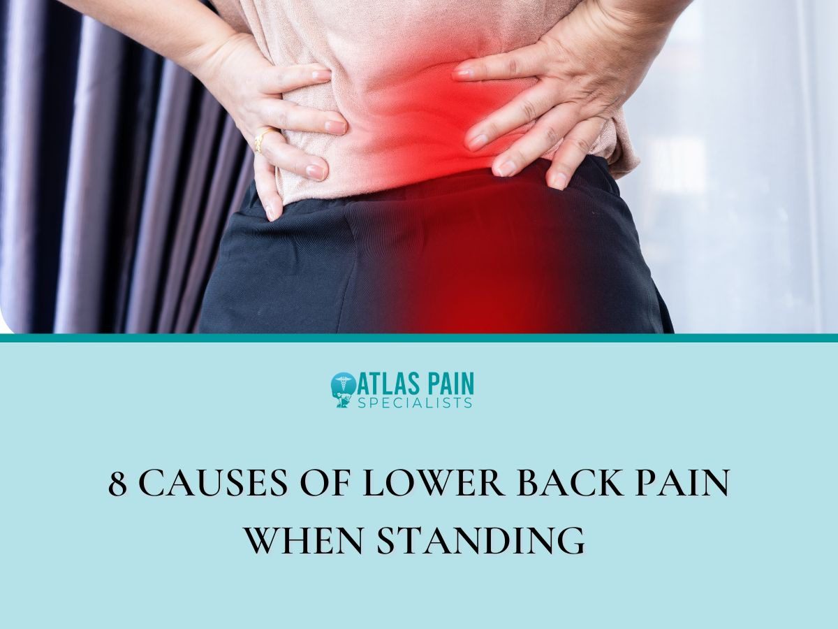
8 Causes of Lower Back Pain When Standing
Date: June 24, 2026
Lower back pain during standing presents a common clinical complaint that often stems from mechanical stress on the spinal column. The pain emerges from a complex interaction between muscle endurance, joint integrity, and neural structures.
Many individuals find that what begins as a mild ache gradually intensifies the longer they remain upright. This progression provides a critical diagnostic clue for healthcare providers.
The causes range from simple muscle fatigue to degenerative conditions of the spine. Each of the eight distinct mechanisms discussed here offers a pathway toward targeted treatment and prevention strategies. Let's look at 8 causes of lower back pain when standing.
1. Muscle Fatigue and Poor Endurance
Sustained standing forces the paraspinal muscles to contract continuously against gravity. Weakness in the abdominal and gluteal muscles compounds this workload because the erector spinae must compensate for insufficient core support.
Fatigue Onset and Core Instability
The first sign of muscle fatigue is often a dull ache that worsens over time. This discomfort develops as metabolic byproducts accumulate within the muscle tissue.
Core musculature functions as a natural corset that stabilizes the lumbar spine. Inadequate core strength permits excessive anterior pelvic tilt during standing, which shifts the center of gravity forward and forces the lower back muscles to work harder.
Endurance and Clinical Assessment
Low endurance in the back extensors predicts earlier onset of standing-related pain. Individuals with sedentary occupations often display reduced paraspinal endurance and remain vulnerable to discomfort during routine standing tasks.
Healthcare providers evaluate muscle fatigue through endurance tests such as the Biering-Sørensen test. A short hold time indicates poor extensor endurance and suggests a need for targeted strengthening.
2. A Pelvis That Tilts Too Far Forward
Anterior pelvic tilt describes a postural deviation where the front of the pelvis drops downward and the rear elevates. This position increases the lumbar lordosis curve and places excessive compression on the facet joints of the lower spine.
The muscle imbalance responsible for this tilt involves tight hip flexors and weak hamstrings. The erector spinae muscles contract forcefully to counterbalance the forward pull of the pelvis, which leads to chronic strain.
The Postural Mechanism
The iliopsoas muscle shortens in individuals who sit for prolonged periods. This shortened position pulls the pelvis into an anterior tilt whenever the person stands.
The abdominal muscles become lengthened and weakened in this posture. Their reduced ability to oppose the hip flexors permits the tilt to persist throughout standing activities.
Pain Distribution and Symptoms
Pain from anterior pelvic tilt localizes to the lower lumbar region and the sacroiliac joints. The ache often presents as a deep, diffuse discomfort rather than a sharp, localized sensation.
Patients frequently report increased pain at the end of the day. This pattern reflects the cumulative loading on spinal structures over hours of standing.
Correction Strategies
Stretching the hip flexors and quadriceps reduces the anterior pull on the pelvis. Strengthening the gluteal muscles provides the posterior counterforce needed to restore neutral alignment.
A physical therapist can assess pelvic position during standing and sitting. Treatment typically includes a home exercise program focused on these specific muscle groups.
3. A Misaligned Pelvis and Postural Habits
Swayback posture differs from anterior pelvic tilt in that the entire pelvis shifts forward relative to the feet. The upper body leans backward to maintain balance, which creates a straight but slouched spinal alignment.
This posture places excessive stress on the posterior ligaments of the lumbar spine. The ligaments stretch over time and lose their ability to provide passive support during standing.
The Swayback Mechanism
The hip joints extend and the thoracic spine moves posteriorly in this stance. The lower back flattens rather than curves excessively, which redistributes load to the connective tissues.
The hamstrings and gluteal muscles remain in a lengthened and weakened state. Their reduced activation forces the lumbar ligaments and joint capsules to bear more of the gravitational load.
Pain Characteristics
Pain from swayback posture typically presents as a chronic ache across the lower back. The discomfort intensifies after fifteen to twenty minutes of quiet standing.
Patients often report that shifting weight from one foot to the other provides temporary relief. This movement reduces ligamentous tension and redistributes compressive forces across the spine.
Habitual Factors and Correction
Prolonged standing on soft surfaces or with poor footwear contributes to this postural pattern. The lack of proprioceptive feedback from the feet allows the pelvis to drift forward without conscious correction.
Re-education of standing posture focuses on weight distribution over the midfoot. Training the gluteal muscles to engage during standing helps anchor the pelvis in a more stable position.
4. A Spinal Disc Issue
Intervertebral discs serve as shock absorbers between the vertebral bodies. Each disc contains a gelatinous nucleus pulposus surrounded by a tough annulus fibrosus.
Standing increases the hydrostatic pressure within the disc's nucleus. This pressure can cause the annulus to bulge outward, particularly when the spine is not in a neutral position.
Bulging and Herniation Mechanics
A bulging disc protrudes beyond its normal boundaries but remains intact. A herniated disc involves a tear in the annulus that allows the nucleus to escape outward.
The direction of the protrusion determines the symptoms. A posterolateral bulge often contacts the spinal nerve root and produces radiating pain.
Pain Patterns and Aggravating Factors
Discogenic pain typically worsens with prolonged standing and improves with lying flat. The reduction in axial loading when supine decreases pressure on the disc and relieves nerve irritation.
Forward bending while standing further compresses the anterior disc. This compression can exacerbate a pre-existing bulge or herniation.
Diagnostic and Treatment Approaches
Magnetic resonance imaging provides the most detailed view of disc morphology. This imaging identifies the location and extent of any disc abnormality.
Conservative treatment includes physical therapy and anti-inflammatory medications. Severe cases may require epidural steroid injections or surgical intervention.
5. The Sciatic Nerve Connection
The sciatic nerve originates from the L4 through S3 nerve roots in the lower spine. This nerve travels through the pelvis and down the posterior aspect of each leg.
Compression or irritation of the sciatic nerve produces pain that radiates along its pathway. Standing can exacerbate this compression by narrowing the intervertebral foramina through which the nerve roots exit.
Mechanisms of Nerve Compression
A herniated disc, bone spur, or narrowed spinal canal can impinge on the sciatic nerve roots. The piriformis muscle in the buttock can also compress the nerve as it passes underneath or through the muscle.
The position of standing increases lumbar extension in some individuals. This extension reduces the space available for the nerve roots within the neural foramina.
Symptom Presentation
Sciatic pain presents as a sharp, shooting sensation that travels from the lower back into the buttock and leg. The pain may be accompanied by tingling, numbness, or a burning sensation in the affected leg.
Symptoms often worsen with standing and improve with sitting or lying down. Flexion of the spine, such as leaning forward, can reduce the nerve compression and provide relief.
Clinical Evaluation
The straight leg raise test helps identify sciatic nerve irritation. This test involves passive elevation of the straightened leg while the patient lies supine.
A positive test reproduces radiating pain down the leg and suggests nerve root involvement. Imaging studies confirm the anatomical source of compression and guide treatment decisions.
6. A Problem With the Facet Joints
Facet joints are paired structures located at the posterior aspect of each vertebral segment. These joints guide spinal motion and provide stability during flexion and extension movements.
Standing in an extended position compresses these joints against each other. This compression generates pain through mechanical stress and local inflammation.
Facet Joint Mechanics
Each facet joint consists of articular cartilage and a synovial lining. The joint capsules contain nerve endings that signal pain when stretched or compressed.
Excessive lumbar lordosis increases the load on the facet joints. This load can cause synovial impingement and capsular irritation over time.
Pain Characteristics
Facet joint pain presents as a deep ache localized to one or both sides of the lumbar spine. The pain does not typically radiate into the leg but may refer to the buttock or thigh.
Extension activities such as standing or walking uphill worsen the symptoms. Flexion of the spine, such as bending forward, provides relief by unloading the joints.
Diagnostic Procedures
Diagnostic facet joint blocks confirm the joint as the pain source. These injections contain local anesthetic placed directly into the joint capsule.
A positive response to the block indicates that the facet joint contributes to the patient's pain. Treatment includes physical therapy, anti-inflammatory medications, and radiofrequency ablation in chronic cases.
7. Spinal Stenosis and Narrowing Space
Spinal stenosis refers to a reduction in the available space within the spinal canal or neural foramina. This narrowing can compress the spinal cord or nerve roots and produce symptoms during standing.
The degenerative form of stenosis results from hypertrophic changes in the facet joints and ligamentum flavum. These structures thicken with age and encroach upon the neural elements.
Central Canal Stenosis
Central stenosis affects the main spinal canal and its contents. Standing with the spine in extension reduces the canal's diameter and worsens nerve compression.
Patients with central stenosis often experience bilateral leg symptoms. These symptoms include pain, weakness, and a heavy sensation in the lower extremities.
Lateral Recess Stenosis
Lateral recess stenosis involves narrowing of the side pockets of the spinal canal. This condition affects the exiting nerve roots as they traverse the lateral recess.
Symptoms present unilaterally and correspond to the specific nerve root affected. The L4 and L5 roots are most frequently involved.
Relief Through Flexion
Patients with stenosis frequently report relief when leaning forward over a shopping cart. This posture increases the spinal canal diameter by creating spinal flexion.
Sitting also reduces symptoms by unloading the spine and opening the canal space. These positional changes serve as important diagnostic clues.
8. Ankylosing Spondylitis and Inflammation
Ankylosing spondylitis is a chronic inflammatory condition that primarily affects the sacroiliac joints and the spine. This autoimmune disease causes enthesitis, which is inflammation at the sites where ligaments and tendons attach to bone.
Standing places axial load on inflamed spinal structures and exacerbates the pain. The inflammatory process leads to progressive stiffness and eventual bony fusion of the vertebral segments.
Disease Mechanism and Onset
The condition has a strong genetic association with the HLA-B27 antigen. This genetic marker appears in over ninety percent of affected individuals.
Inflammation begins at the sacroiliac joints and ascends the spine over years. New bone formation occurs at the margins of the vertebral bodies and produces syndesmophytes.
Pain Characteristics
Inflammatory back pain differs from mechanical back pain in several ways. The pain improves with physical activity and worsens with prolonged rest or stillness.
Morning stiffness lasts longer than thirty minutes after waking. Standing for extended periods triggers deep pain in the lower back and buttocks.
Diagnostic and Treatment Approach
Diagnosis relies on radiographic evidence of sacroiliitis and clinical criteria. Elevated inflammatory markers such as erythrocyte sedimentation rate support the diagnosis.
Treatment includes nonsteroidal anti-inflammatory drugs and biologic agents. Physical therapy maintains spinal mobility and delays the progression of fusion.
Conclusion
Lower back pain during standing arises from a diverse set of mechanical, degenerative, and inflammatory processes. Accurate diagnosis requires a thorough clinical history and physical examination to distinguish between these distinct etiologies.
Self-treatment without professional guidance carries the risk of worsening the underlying condition. A qualified healthcare provider can perform specific tests to isolate the pain source and recommend appropriate interventions.
Treatment strategies range from postural re-education and targeted strengthening to pharmacological management and surgical options. Patients who experience persistent or progressive symptoms should seek medical evaluation without delay.
About Dr. Sean Ormond

Dr. Sean Ormond is dual board-certified in Anesthesiology and Interventional Pain Management. He completed his anesthesia residency at Case Western University in Cleveland, Ohio where he served as Chief Resident, followed by an interventional pain management fellowship at Rush University in Chicago, IL. Following fellowship, Dr. Ormond moved to Phoenix and has been practicing in the Valley for a few years before deciding to start his own practice.