New Location at 8406 E. Shea Blvd #100, Scottsdale 85260 - Accepting New Patients
Pain on Medial Side of Knee: Causes and Relief Options
Date: October 21, 2025
Pain on the inner side of the knee can range from a mild ache after activity to sharp discomfort that limits movement. This area, known as the medial knee region, includes several structures that absorb force, stabilize motion, and bear significant body weight.
When any of these tissues become irritated, injured, or degenerated, even simple movements like climbing stairs or getting out of a chair can cause distress. Persistent discomfort often reflects deeper mechanical or inflammatory problems that deserve careful attention.
Accurate assessment of medial knee pain helps determine whether it stems from joint wear, ligament strain, or inflammation of nearby soft tissues. With precise diagnosis and targeted management, most individuals regain comfort and prevent chronic complications.
Causes Of Pain On Medial Side Of Knee
The medial knee houses ligaments, cartilage, tendons, and bursae that each play a role in stability and load transfer. When one of these components becomes compromised, pain tends to localize sharply along the inner joint line.

| Cause | Description | Typical Triggers or Risk Factors | Pain Characteristics |
| MCL Injury | Stretching or tearing of the inner stabilizing ligament. | Sports impact, sudden twists. | Sharp medial pain, swelling, instability. |
| Meniscus Tear | Damage to inner cartilage cushion. | Deep bending, pivoting under load. | Clicking, catching, pain during rotation. |
| Pes Anserine Bursitis | Inflammation of bursa below joint. | Overuse, tight hamstrings, obesity. | Local tenderness below the joint line. |
| Osteoarthritis | Cartilage thinning in the medial compartment. | Aging, repetitive load, past trauma. | Dull ache, stiffness, morning discomfort. |
| Medial Plica Syndrome | Irritation of inner knee fold. | Repetitive flexion, overuse. | Snapping, mild swelling, intermittent pain. |
Common causes include acute injuries such as ligament sprains and cartilage tears, as well as gradual conditions like osteoarthritis and bursitis. Each presents distinct symptoms and treatment priorities depending on the underlying tissue involved.
- Medial Collateral Ligament (MCL) Injury
The medial collateral ligament stabilizes the inner knee against sideways pressure. Sudden twisting or direct blows during sports frequently strain or tear this ligament.
Pain from an MCL injury appears immediately after the incident and may worsen with sideward motion. Mild sprains often improve with rest, while severe tears sometimes require bracing or rehabilitation guided by a physiotherapist.
A diagnostic examination usually includes the valgus stress test, where outward pressure on the knee reproduces pain if the MCL is compromised. Recovery depends on injury grade, with early protection and progressive strengthening supporting full return to activity.
- Medial Meniscus Tear
The meniscus is a cartilage disc that cushions and stabilizes the knee joint. The medial meniscus endures higher forces than the outer counterpart, making it prone to tears.
Degenerative wear or a sudden twist while bearing weight can cause the cartilage to tear, producing sharp pain and occasional locking. Symptoms often worsen with squatting, pivoting, or prolonged standing.
Diagnosis may involve MRI imaging to confirm the tear pattern and extent. Treatment ranges from physiotherapy and anti-inflammatory measures to arthroscopic repair in cases with mechanical blockage.
- Pes Anserine Bursitis
Below the joint line lies a small fluid-filled sac called the pes anserine bursa. Its purpose is to reduce friction between tendons and the bone during knee movement.
Repetitive stress, obesity, or tight hamstrings can inflame this bursa, leading to localized swelling and pain below the inner knee. Tenderness usually intensifies during stair climbing or side-lying positions at night.
Initial management involves rest, ice application, and targeted stretching to reduce friction on the affected area. If symptoms persist, corticosteroid injections or physiotherapy focusing on muscle balance may be considered.
Key Symptoms and Diagnostic Clues
Medial knee pain can mimic multiple conditions that share overlapping sensations. Differentiating among them relies on precise clinical evaluation and targeted testing.
Doctors assess joint alignment, palpate the tender region, and may perform movement-based tests to reproduce pain. Imaging such as X-rays or MRI clarifies structural changes when physical signs are inconclusive.
Characteristic Pain Patterns
Pain at the inner joint line often reflects cartilage or ligament injury. Sharp localized tenderness indicates mechanical involvement rather than diffuse inflammation.
Patients sometimes report a catching or locking feeling when a meniscus tear interferes with smooth motion. Swelling limited to the inner region commonly suggests bursitis or minor sprain rather than generalized joint fluid buildup.
Associated stiffness after inactivity typically accompanies degenerative processes like osteoarthritis. These observations guide clinicians toward more precise testing before intervention begins.
| Indicator | Possible Cause | Notes |
| Pain on twisting | Meniscus tear | Often produces clicking or locking. |
| Tenderness on inner joint line | Osteoarthritis or meniscus lesion | Worsens after activity. |
| Pain below joint line | Pes anserine bursitis | Increases with climbing or kneeling. |
| Valgus stress reproduces pain | MCL injury | Confirms ligament instability. |
| Stiffness easing with motion | Osteoarthritis | Indicates chronic cartilage wear. |
Relief and Treatment
Effective treatment depends on cause, severity, and the patient’s activity level. Most cases improve with non-surgical interventions emphasizing rest, strength, and controlled movement.
Doctors often begin with conservative management before considering injections or surgery. The goal is to relieve discomfort while preserving long-term knee function.
- Home and Conservative Care
Initial relief often involves the RICE protocol: rest, ice, compression, and elevation. These measures reduce inflammation and limit swelling in the early stages.
Supportive braces may stabilize the knee and prevent excessive side movement during recovery. Over-the-counter anti-inflammatory medication can help ease pain in mild injuries.
Consider:
- Limiting activities that cause twisting or sideward strain.
- Applying ice for 15–20 minutes several times daily.
- Using a soft brace if recommended by a professional.
- Avoiding prolonged immobilization to prevent stiffness.
- Physical Therapy and Exercise
Therapeutic programs aim to restore strength, flexibility, and joint alignment. Exercises typically focus on quadriceps, hamstrings, and hip stability.
A physiotherapist tailors routines to avoid aggravating sensitive areas. Gradual load progression ensures tissue adaptation without overstrain.
Popular exercises include wall sits, hamstring curls, and step-ups under professional supervision. Balance training also enhances proprioception, lowering reinjury risk once normal activity resumes.
- Medical and Surgical Interventions
When conservative care fails, physicians may recommend targeted injections or minimally invasive surgery. These options are reserved for persistent or structural causes of pain.
| Approach | Purpose | Examples |
| Rest and Load Control | Reduces stress on healing tissue. | Short rest followed by gradual activity. |
| Ice and Compression | Limits inflammation. | 15–20 minutes several times per day. |
| Physical Therapy | Restores motion and strength. | Muscle balancing, flexibility work. |
| Bracing or Taping | Improves stability. | Hinged braces for MCL support. |
| Medication | Eases pain and swelling. | NSAIDs, topical gels. |
| Injections | Reduces chronic inflammation. | Corticosteroid or hyaluronic acid. |
| Surgery | Repairs structural damage. | Meniscus repair or realignment. |
Corticosteroid injections can temporarily reduce inflammation in bursitis or arthritis. Hyaluronic acid may improve joint lubrication and mobility in degenerative cases.
Surgical repair or removal of torn meniscus fragments is considered when locking or mechanical instability interferes with daily function. Early rehabilitation after surgery preserves joint motion and strength.
How to Prevent Future Knee Pain
Preventing medial knee pain involves maintaining strength, flexibility, and proper movement mechanics. These measures reduce repetitive strain on vulnerable inner structures.
Lifestyle choices like balanced exercise routines and joint-friendly footwear contribute significantly to protection. Consistency in care yields lasting comfort and mobility.
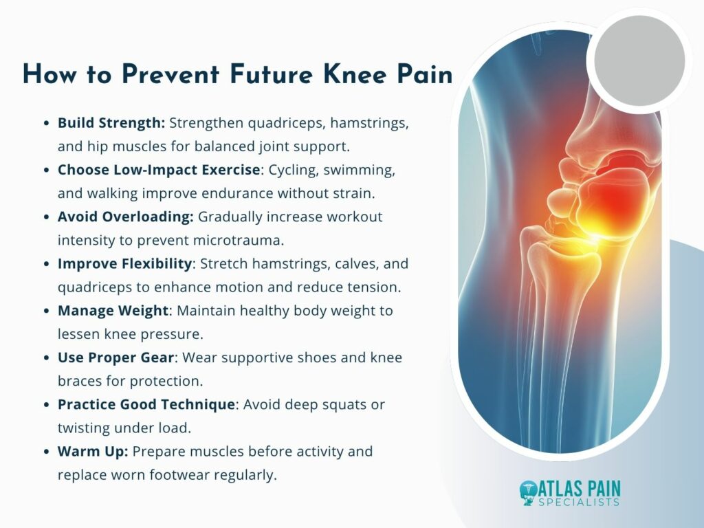
- Strength and Conditioning
Strong muscles support and offload stress from the knee joint. Balanced training of quadriceps, hamstrings, and hip abductors provides symmetrical control.
Low-impact activities such as cycling, swimming, and brisk walking build endurance without overloading the joint. Avoiding sudden intensity spikes helps prevent recurrent microtrauma.
Regular assessment of movement form ensures alignment during both exercise and daily tasks. Even minor posture corrections can meaningfully reduce medial strain.
- Flexibility and Weight Control
Tight muscles alter knee mechanics and contribute to bursitis or ligament tension. Stretching of hamstrings, calves, and quadriceps enhances range of motion.
Weight management lowers compressive forces that accelerate cartilage wear. A moderate calorie intake with nutrient-rich food supports overall joint health.
Professional guidance from a physiotherapist or trainer ensures proper technique and progress monitoring. Sustainable habits outperform occasional corrective efforts.
- Protective Equipment and Technique
Athletes and workers benefit from using knee supports when engaging in repetitive or high-impact tasks. Proper footwear with supportive midsoles maintains leg alignment.
Avoiding deep squats or twisting under heavy load prevents strain on ligaments and cartilage. Gradual warm-up routines prepare soft tissues for movement.
Remember:
- Warm up before intense activity.
- Replace worn footwear regularly.
- Maintain even leg strength and posture awareness.
When to Seek Medical Evaluation
Ongoing pain on the inner side of the knee should not be dismissed as a simple strain or temporary soreness. Medical evaluation helps identify tissue injury, inflammation, or early degeneration before these issues worsen.
Even mild symptoms can mask damage to ligaments, cartilage, or the joint lining. Timely consultation with a healthcare provider ensures that the underlying cause receives appropriate and precise treatment.
- Identify Signs Early
Certain symptoms indicate a need for immediate medical attention. Sudden swelling, bruising, or a sensation of tearing often points to meniscus or ligament injury.
A popping sound during motion or loss of stability suggests structural disruption inside the joint. Difficulty bearing weight or locking during flexion may signal mechanical obstruction from loose fragments or displaced tissue.
Redness, warmth, or fever around the knee can imply infection or inflammatory disease. In these cases, prompt diagnosis prevents joint damage and reduces the risk of long-term complications.
- When Chronic Pain Persists
Pain that lasts longer than two weeks or worsens with activity can indicate degenerative or inflammatory processes. Osteoarthritis, tendinopathy, and synovial irritation commonly present with dull, recurring discomfort.
Morning stiffness, grinding sensations, or clicking sounds often accompany cartilage thinning or irregular joint surfaces. Without timely management, these mechanical changes may limit range of motion and increase fatigue during walking or climbing.
Recurrent swelling after moderate exertion suggests underlying joint instability or poor alignment. Continuous irritation in the same region warrants orthopedic review for targeted treatment and corrective exercises.
- Diagnostic Steps and Professional Assessment
Physicians begin by evaluating movement patterns, palpating the joint, and testing flexibility. These manual assessments provide essential clues about which structures contribute to pain.
Imaging tests like X-rays, ultrasound, or MRI reveal the extent of tissue injury or degeneration. MRI scans are especially useful for detecting meniscal tears, cartilage wear, and inflammation around tendons or ligaments.
Blood tests may accompany imaging if infection or systemic inflammation is suspected. Identifying biochemical markers helps guide medication and rehabilitation decisions.
Benefits of Early Medical Intervention
Prompt evaluation enables precise diagnosis and prevents mild symptoms from escalating into chronic conditions. Early treatment often limits inflammation and protects surrounding tissues from overcompensation strain.
Professional care helps establish structured recovery through physical therapy and supervised exercise. This approach reduces pain while restoring coordination, flexibility, and confidence in movement.
Ignoring early warning signs can extend recovery time and increase the likelihood of reinjury. Seeking timely medical input keeps the healing process efficient and minimizes long-term joint stress.
Hurts to Straighten Knee? Here's What You Need To Know
Experiencing inner knee pain shows how balance, alignment, and tissue resilience determine overall joint comfort. Careful evaluation and early treatment can restore smooth motion and prevent recurring discomfort.
As pain subsides, attention to movement quality and muscular stability becomes essential for long-term relief. Consistent rehabilitation and joint-friendly activity preserve strength while protecting the inner structures of the knee.
Effective recovery depends on sustained awareness of daily movement habits and posture. When managed thoughtfully, the knee regains stability, allowing confidence in movement without lingering pain.
About Dr. Sean Ormond

Dr. Sean Ormond is dual board-certified in Anesthesiology and Interventional Pain Management. He completed his anesthesia residency at Case Western University in Cleveland, Ohio where he served as Chief Resident, followed by an interventional pain management fellowship at Rush University in Chicago, IL. Following fellowship, Dr. Ormond moved to Phoenix and has been practicing in the Valley for a few years before deciding to start his own practice.